+ データを開く
- 基本情報
基本情報
| 登録情報 | データベース: EMDB / ID: EMD-13760 | |||||||||
|---|---|---|---|---|---|---|---|---|---|---|
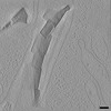
| タイトル | P. carterae coccolith vesicle with growing crystals | |||||||||
 マップデータ マップデータ | P. carterae coccolith vesicle with growing crystals | |||||||||
 試料 試料 |
| |||||||||
| 生物種 |  Chrysotila carterae (真核生物) Chrysotila carterae (真核生物) | |||||||||
| 手法 | 電子線トモグラフィー法 / クライオ電子顕微鏡法 | |||||||||
 データ登録者 データ登録者 | Kadan Y / Mahamid J / Gal A / Tollervey F | |||||||||
| 資金援助 | European Union, 2件
| |||||||||
 引用 引用 |  ジャーナル: Proc Natl Acad Sci U S A / 年: 2021 ジャーナル: Proc Natl Acad Sci U S A / 年: 2021タイトル: Intracellular nanoscale architecture as a master regulator of calcium carbonate crystallization in marine microalgae. 著者: Yuval Kadan / Fergus Tollervey / Neta Varsano / Julia Mahamid / Assaf Gal /   要旨: Unicellular marine microalgae are responsible for one of the largest carbon sinks on Earth. This is in part due to intracellular formation of calcium carbonate scales termed coccoliths. ...Unicellular marine microalgae are responsible for one of the largest carbon sinks on Earth. This is in part due to intracellular formation of calcium carbonate scales termed coccoliths. Traditionally, the influence of changing environmental conditions on this process has been estimated using poorly constrained analogies to crystallization mechanisms in bulk solution, yielding ambiguous predictions. Here, we elucidated the intracellular nanoscale environment of coccolith formation in the model species using cryoelectron tomography. By visualizing cells at various stages of the crystallization process, we reconstructed a timeline of coccolith development. The three-dimensional data portray the native-state structural details of coccolith formation, uncovering the crystallization mechanism, and how it is spatially and temporally controlled. Most strikingly, the developing crystals are only tens of nanometers away from delimiting membranes, resulting in a highly confined volume for crystal growth. We calculate that the number of soluble ions that can be found in such a minute volume at any given time point is less than the number needed to allow the growth of a single atomic layer of the crystal and that the uptake of single protons can markedly affect nominal pH values. In such extreme confinement, the crystallization process is expected to depend primarily on the regulation of ion fluxes by the living cell, and nominal ion concentrations, such as pH, become the result, rather than a driver, of the crystallization process. These findings call for a new perspective on coccolith formation that does not rely exclusively on solution chemistry. | |||||||||
| 履歴 |
|
- 構造の表示
構造の表示
| ムービー |
 ムービービューア ムービービューア |
|---|---|
| 添付画像 |
- ダウンロードとリンク
-EMDBアーカイブ
| マップデータ |  emd_13760.map.gz emd_13760.map.gz | 315.9 MB |  EMDBマップデータ形式 EMDBマップデータ形式 | |
|---|---|---|---|---|
| ヘッダ (付随情報) |  emd-13760-v30.xml emd-13760-v30.xml emd-13760.xml emd-13760.xml | 10.1 KB 10.1 KB | 表示 表示 |  EMDBヘッダ EMDBヘッダ |
| 画像 |  emd_13760.png emd_13760.png | 204.1 KB | ||
| アーカイブディレクトリ |  http://ftp.pdbj.org/pub/emdb/structures/EMD-13760 http://ftp.pdbj.org/pub/emdb/structures/EMD-13760 ftp://ftp.pdbj.org/pub/emdb/structures/EMD-13760 ftp://ftp.pdbj.org/pub/emdb/structures/EMD-13760 | HTTPS FTP |
-検証レポート
| 文書・要旨 |  emd_13760_validation.pdf.gz emd_13760_validation.pdf.gz | 297.3 KB | 表示 |  EMDB検証レポート EMDB検証レポート |
|---|---|---|---|---|
| 文書・詳細版 |  emd_13760_full_validation.pdf.gz emd_13760_full_validation.pdf.gz | 296.9 KB | 表示 | |
| XML形式データ |  emd_13760_validation.xml.gz emd_13760_validation.xml.gz | 3.7 KB | 表示 | |
| CIF形式データ |  emd_13760_validation.cif.gz emd_13760_validation.cif.gz | 4.6 KB | 表示 | |
| アーカイブディレクトリ |  https://ftp.pdbj.org/pub/emdb/validation_reports/EMD-13760 https://ftp.pdbj.org/pub/emdb/validation_reports/EMD-13760 ftp://ftp.pdbj.org/pub/emdb/validation_reports/EMD-13760 ftp://ftp.pdbj.org/pub/emdb/validation_reports/EMD-13760 | HTTPS FTP |
-関連構造データ
- リンク
リンク
| EMDBのページ |  EMDB (EBI/PDBe) / EMDB (EBI/PDBe) /  EMDataResource EMDataResource |
|---|
- マップ
マップ
| ファイル |  ダウンロード / ファイル: emd_13760.map.gz / 形式: CCP4 / 大きさ: 821.3 MB / タイプ: IMAGE STORED AS SIGNED INTEGER (2 BYTES) ダウンロード / ファイル: emd_13760.map.gz / 形式: CCP4 / 大きさ: 821.3 MB / タイプ: IMAGE STORED AS SIGNED INTEGER (2 BYTES) | ||||||||||||||||||||||||||||||||||||||||||||||||||||||||||||
|---|---|---|---|---|---|---|---|---|---|---|---|---|---|---|---|---|---|---|---|---|---|---|---|---|---|---|---|---|---|---|---|---|---|---|---|---|---|---|---|---|---|---|---|---|---|---|---|---|---|---|---|---|---|---|---|---|---|---|---|---|---|
| 注釈 | P. carterae coccolith vesicle with growing crystals | ||||||||||||||||||||||||||||||||||||||||||||||||||||||||||||
| 投影像・断面図 | 画像のコントロール
画像は Spider により作成 これらの図は立方格子座標系で作成されたものです | ||||||||||||||||||||||||||||||||||||||||||||||||||||||||||||
| ボクセルのサイズ | X=Y=Z: 13.48 Å | ||||||||||||||||||||||||||||||||||||||||||||||||||||||||||||
| 密度 |
| ||||||||||||||||||||||||||||||||||||||||||||||||||||||||||||
| 対称性 | 空間群: 1 | ||||||||||||||||||||||||||||||||||||||||||||||||||||||||||||
| 詳細 | EMDB XML:
CCP4マップ ヘッダ情報:
| ||||||||||||||||||||||||||||||||||||||||||||||||||||||||||||
-添付データ
- 試料の構成要素
-全体 : Pleurochrysis carterae
| 全体 | 名称: Pleurochrysis carterae |
|---|---|
| 要素 |
|
-超分子 #1: Pleurochrysis carterae
| 超分子 | 名称: Pleurochrysis carterae / タイプ: cell / ID: 1 / 親要素: 0 |
|---|---|
| 由来(天然) | 生物種:  Chrysotila carterae (真核生物) Chrysotila carterae (真核生物) |
-実験情報
-構造解析
| 手法 | クライオ電子顕微鏡法 |
|---|---|
 解析 解析 | 電子線トモグラフィー法 |
| 試料の集合状態 | cell |
- 試料調製
試料調製
| 緩衝液 | pH: 8 詳細: Sterile artificial seawater, supplemented with an f/2 nutrient recipe |
|---|---|
| グリッド | モデル: Quantifoil R2/1 / 材質: COPPER / メッシュ: 200 / 支持フィルム - 材質: CARBON / 支持フィルム - トポロジー: HOLEY / 前処理 - タイプ: PLASMA CLEANING / 前処理 - 雰囲気: AIR / 前処理 - 気圧: 0.037 kPa / 詳細: 15 mA |
| 凍結 | 凍結剤: ETHANE / 装置: LEICA EM GP |
| 切片作成 | 集束イオンビーム - 装置: OTHER / 集束イオンビーム - イオン: OTHER / 集束イオンビーム - 電圧: 30 kV / 集束イオンビーム - 電流: 0.05 nA / 集束イオンビーム - 時間: 1800 sec. / 集束イオンビーム - 温度: 90 K / 集束イオンビーム - Initial thickness: 5000 nm / 集束イオンビーム - 最終 厚さ: 200 nm 集束イオンビーム - 詳細: For details, see publication. The value given for _emd_sectioning_focused_ion_beam.instrument is FEI Aquilos. This is not in a list of allowed values {'DB235', ...集束イオンビーム - 詳細: For details, see publication. The value given for _emd_sectioning_focused_ion_beam.instrument is FEI Aquilos. This is not in a list of allowed values {'DB235', 'OTHER'} so OTHER is written into the XML file. |
- 電子顕微鏡法
| 顕微鏡 | FEI TITAN KRIOS |
|---|---|
| 特殊光学系 | 位相板: VOLTA PHASE PLATE / エネルギーフィルター - 名称: GIF Quantum LS / エネルギーフィルター - スリット幅: 20 eV |
| 撮影 | フィルム・検出器のモデル: GATAN K2 SUMMIT (4k x 4k) 検出モード: COUNTING / デジタル化 - サイズ - 横: 3710 pixel / デジタル化 - サイズ - 縦: 3838 pixel / 平均露光時間: 1.6 sec. / 平均電子線量: 2.24 e/Å2 |
| 電子線 | 加速電圧: 300 kV / 電子線源:  FIELD EMISSION GUN FIELD EMISSION GUN |
| 電子光学系 | 照射モード: FLOOD BEAM / 撮影モード: BRIGHT FIELD / Cs: 2.7 mm / 倍率(公称値): 42000 |
| 試料ステージ | 試料ホルダーモデル: FEI TITAN KRIOS AUTOGRID HOLDER ホルダー冷却材: NITROGEN |
| 実験機器 |  モデル: Titan Krios / 画像提供: FEI Company |
- 画像解析
画像解析
| 最終 再構成 | ソフトウェア - 名称:  IMOD / 使用した粒子像数: 50 IMOD / 使用した粒子像数: 50 |
|---|
 ムービー
ムービー コントローラー


 Z (Sec.)
 Y (Row.)
 X (Col.)


