 ムービー
ムービー コントローラー


+ データを開く

- 基本情報
基本情報
| 登録情報 | データベース: EMDB / ID: EMD-13672 | ||||||||||||||||||
|---|---|---|---|---|---|---|---|---|---|---|---|---|---|---|---|---|---|---|---|
| タイトル | Sub-tomogram average of Ca. M.lanthanidiphila S-layer obtained from whole cell cryo-tomograms | ||||||||||||||||||
 マップデータ マップデータ | Sub-tomogram average of the S-layer of Ca. M.lanthanidiphila obtained from whole cell cryo-tomograms | ||||||||||||||||||
 試料 試料 |
| ||||||||||||||||||
 キーワード キーワード | S-layer / STRUCTURAL PROTEIN | ||||||||||||||||||
| 生物種 |  Candidatus Methylomirabilis lanthanidiphila (バクテリア) Candidatus Methylomirabilis lanthanidiphila (バクテリア) | ||||||||||||||||||
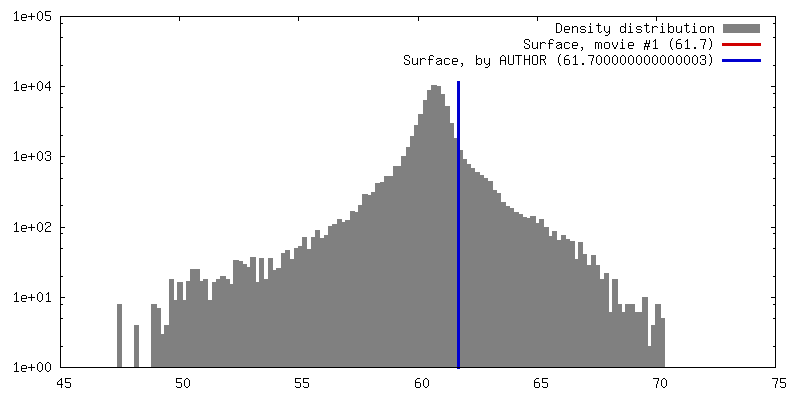
| 手法 | サブトモグラム平均法 / クライオ電子顕微鏡法 / 解像度: 25.0 Å | ||||||||||||||||||
 データ登録者 データ登録者 | Gambelli L / Mesman R | ||||||||||||||||||
| 資金援助 | European Union, 5件
| ||||||||||||||||||
 引用 引用 | ジャーナル: Front Microbiol / 年: 2021 タイトル: The Polygonal Cell Shape and Surface Protein Layer of Anaerobic Methane-Oxidizing Bacteria. 著者: Lavinia Gambelli / Rob Mesman / Wouter Versantvoort / Christoph A Diebolder / Andreas Engel / Wiel Evers / Mike S M Jetten / Martin Pabst / Bertram Daum / Laura van Niftrik /   要旨: bacteria perform anaerobic methane oxidation coupled to nitrite reduction via an intra-aerobic pathway, producing carbon dioxide and dinitrogen gas. These diderm bacteria possess an unusual ... bacteria perform anaerobic methane oxidation coupled to nitrite reduction via an intra-aerobic pathway, producing carbon dioxide and dinitrogen gas. These diderm bacteria possess an unusual polygonal cell shape with sharp ridges that run along the cell body. Previously, a putative surface protein layer (S-layer) was observed as the outermost cell layer of these bacteria. We hypothesized that this S-layer is the determining factor for their polygonal cell shape. Therefore, we enriched the S-layer from cells and through LC-MS/MS identified a 31 kDa candidate S-layer protein, mela_00855, which had no homology to any other known protein. Antibodies were generated against a synthesized peptide derived from the mela_00855 protein sequence and used in immunogold localization to verify its identity and location. Both on thin sections of cells and in negative-stained enriched S-layer patches, the immunogold localization identified mela_00855 as the S-layer protein. Using electron cryo-tomography and sub-tomogram averaging of S-layer patches, we observed that the S-layer has a hexagonal symmetry. Cryo-tomography of whole cells showed that the S-layer and the outer membrane, but not the peptidoglycan layer and the cytoplasmic membrane, exhibited the polygonal shape. Moreover, the S-layer consisted of multiple rigid sheets that partially overlapped, most likely giving rise to the unique polygonal cell shape. These characteristics make the S-layer of a distinctive and intriguing case to study. | ||||||||||||||||||
| 履歴 |
|
- 構造の表示
構造の表示
| ムービー |
 ムービービューア ムービービューア |
|---|---|
| 構造ビューア | EMマップ: SurfViewMolmilJmol/JSmol |
| 添付画像 |

- ダウンロードとリンク
-EMDBアーカイブ
| マップデータ | emd_13672.map.gz | 201.3 KB | EMDBマップデータ形式 | |
|---|---|---|---|---|
| ヘッダ (付随情報) | emd-13672-v30.xmlemd-13672.xml | 13.1 KB 13.1 KB | 表示 表示 | EMDBヘッダ |
| 画像 | 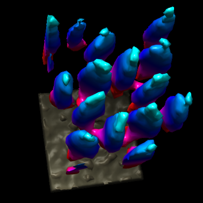 emd_13672.png emd_13672.png | 93.5 KB | ||
| Filedesc metadata | emd-13672.cif.gz | 4.6 KB | ||
| アーカイブディレクトリ |  http://ftp.pdbj.org/pub/emdb/structures/EMD-13672ftp://ftp.pdbj.org/pub/emdb/structures/EMD-13672 http://ftp.pdbj.org/pub/emdb/structures/EMD-13672ftp://ftp.pdbj.org/pub/emdb/structures/EMD-13672 | HTTPS FTP |
-検証レポート
| 文書・要旨 | emd_13672_validation.pdf.gz | 370.5 KB | 表示 | EMDB検証レポート |
|---|---|---|---|---|
| 文書・詳細版 | emd_13672_full_validation.pdf.gz | 370 KB | 表示 | |
| XML形式データ | emd_13672_validation.xml.gz | 4.6 KB | 表示 | |
| CIF形式データ | emd_13672_validation.cif.gz | 5.1 KB | 表示 | |
| アーカイブディレクトリ | https://ftp.pdbj.org/pub/emdb/validation_reports/EMD-13672ftp://ftp.pdbj.org/pub/emdb/validation_reports/EMD-13672 | HTTPS FTP |
-関連構造データ
| 関連構造データ | C: 同じ文献を引用 ( |
|---|---|
| 電子顕微鏡画像生データ | EMPIAR-10829 (タイトル: Cryo Electron Tomography of Ca.M.lanthanidiphila for subtomographic averaging of the S-layer Data size: 135.7 Data #1: Reconstructed tomogram of Ca. M.lanthanidiphila cell [reconstructed volumes] Data #2: Reconstructed tomogram of Ca. M.lanthanidiphila S-layer cell [reconstructed volumes]) |

-リンク
| EMDBのページ | EMDB (EBI/PDBe) / EMDataResource |
|---|
-マップ
| ファイル | ダウンロード / ファイル: emd_13672.map.gz / 形式: CCP4 / 大きさ: 313.5 KB / タイプ: IMAGE STORED AS FLOATING POINT NUMBER (4 BYTES) | ||||||||||||||||||||||||||||||||||||||||||||||||||||||||||||||||||||
|---|---|---|---|---|---|---|---|---|---|---|---|---|---|---|---|---|---|---|---|---|---|---|---|---|---|---|---|---|---|---|---|---|---|---|---|---|---|---|---|---|---|---|---|---|---|---|---|---|---|---|---|---|---|---|---|---|---|---|---|---|---|---|---|---|---|---|---|---|---|
| 注釈 | Sub-tomogram average of the S-layer of Ca. M.lanthanidiphila obtained from whole cell cryo-tomograms | ||||||||||||||||||||||||||||||||||||||||||||||||||||||||||||||||||||
| 投影像・断面図 | 画像のコントロール
画像は Spider により作成 これらの図は立方格子座標系で作成されたものです | ||||||||||||||||||||||||||||||||||||||||||||||||||||||||||||||||||||
| ボクセルのサイズ | X=Y=Z: 7.92 Å | ||||||||||||||||||||||||||||||||||||||||||||||||||||||||||||||||||||
| 密度 |
| ||||||||||||||||||||||||||||||||||||||||||||||||||||||||||||||||||||
| 対称性 | 空間群: 1 | ||||||||||||||||||||||||||||||||||||||||||||||||||||||||||||||||||||
| 詳細 | EMDB XML:
CCP4マップ ヘッダ情報:
| ||||||||||||||||||||||||||||||||||||||||||||||||||||||||||||||||||||
 Z (Sec.)
 Y (Row.)
 X (Col.)


-添付データ
- 試料の構成要素
-全体 : S-layer of Ca.M.lanthanidiphila
| 全体 | 名称: S-layer of Ca.M.lanthanidiphila |
|---|---|
| 要素 |
|
-超分子 #1: S-layer of Ca.M.lanthanidiphila
| 超分子 | 名称: S-layer of Ca.M.lanthanidiphila / タイプ: organelle_or_cellular_component / ID: 1 / 親要素: 0 詳細: 3ml Ca. M.lanthanidiphila floculant biomass was dispersed using a ball bearing homogenizer with 8um clearance. |
|---|---|
| 由来(天然) | 生物種: Candidatus Methylomirabilis lanthanidiphila (バクテリア) 細胞中の位置: on top of outer membrane |
| 分子量 | 理論値: 31.6 KDa |
-実験情報
-構造解析
| 手法 | クライオ電子顕微鏡法 |
|---|---|
 解析 解析 | サブトモグラム平均法 |
| 試料の集合状態 | cell |
-試料調製
| 緩衝液 | pH: 7 |
|---|---|
| グリッド | モデル: Quantifoil R2/2 / 材質: COPPER / メッシュ: 200 / 前処理 - タイプ: GLOW DISCHARGE / 前処理 - 時間: 40 sec. / 前処理 - 雰囲気: AIR / 前処理 - 気圧: 20.0 kPa |
| 凍結 | 凍結剤: ETHANE / チャンバー内湿度: 100 % / チャンバー内温度: 293 K / 装置: FEI VITROBOT MARK IV 詳細: 2 microlitre sample was mixed with 0.5 microlitre 10 nm ProteinA gold solution Grids were plunge frozen in liquid ethane, blot force 1, blot time 3 sec.. |
| 詳細 | Dispersed cells from floculant biomass |
- 電子顕微鏡法
| 顕微鏡 | FEI TITAN KRIOS |
|---|---|
| 温度 | 最高: 93.0 K |
| 特殊光学系 | エネルギーフィルター - 名称: GIF Bioquantum / エネルギーフィルター - スリット幅: 20 eV |
| 撮影 | フィルム・検出器のモデル: GATAN K3 BIOQUANTUM (6k x 4k) デジタル化 - サイズ - 横: 4092 pixel / デジタル化 - サイズ - 縦: 5760 pixel / 撮影したグリッド数: 1 / 平均露光時間: 0.3835 sec. / 平均電子線量: 1.63 e/Å2 |
| 電子線 | 加速電圧: 300 kV / 電子線源:  FIELD EMISSION GUN FIELD EMISSION GUN |
| 電子光学系 | C2レンズ絞り径: 50.0 µm / 最大 デフォーカス(補正後): 4.68 µm / 最小 デフォーカス(補正後): 4.13 µm / 照射モード: FLOOD BEAM / 撮影モード: BRIGHT FIELD / Cs: 2.7 mm / 最大 デフォーカス(公称値): 5.0 µm / 最小 デフォーカス(公称値): 4.0 µm / 倍率(公称値): 33000 |
| 試料ステージ | 試料ホルダーモデル: FEI TITAN KRIOS AUTOGRID HOLDER ホルダー冷却材: NITROGEN |
| 実験機器 |  モデル: Titan Krios / 画像提供: FEI Company |
-画像解析
| 最終 再構成 | 使用したクラス数: 1 / 想定した対称性 - 点群: C6 (6回回転対称) / アルゴリズム: BACK PROJECTION / 解像度のタイプ: BY AUTHOR / 解像度: 25.0 Å / 解像度の算出法: OTHER / ソフトウェア - 名称: PEET (ver. 1.14.0) / 詳細: resolution obtained from FFT of obtainded average / 使用したサブトモグラム数: 1330 |
|---|---|
| 抽出 | トモグラム数: 3 / 使用した粒子像数: 6000 / 参照モデル: averaged from hand picked particles / 手法: random grid / ソフトウェア - 名称: PEET (ver. 1.14.0) 詳細: Used binnend tomograms. Excized sub-volumes of the S-layer with the slicer tool, applied a grid model and saved the grid model after projection on the whole tomogram. |
| 最終 角度割当 | タイプ: NOT APPLICABLE |
