 Movie
Movie Controller


+ Open data

- Basic information
| Entry |  | ||||||||||||
|---|---|---|---|---|---|---|---|---|---|---|---|---|---|
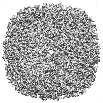
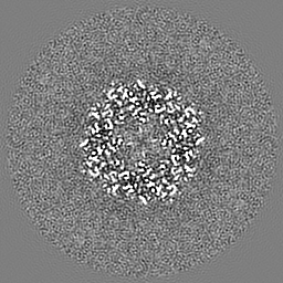
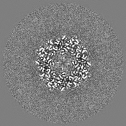
| Title | Horse spleen apoferritin frozen under anaerobic conditions | ||||||||||||
 Map data Map data | |||||||||||||
 Sample Sample |
| ||||||||||||
| Biological species |  | ||||||||||||
| Method | single particle reconstruction / cryo EM / Resolution: 2.4 Å | ||||||||||||
 Authors Authors | Cherrier MV / Vernede X / Fenel D / Martin L / Arragain B / Neumann E / Fontecilla-Camps JC / Schoehn G / Nicolet Y | ||||||||||||
| Funding support |  France, 3 items France, 3 items
| ||||||||||||
 Citation Citation | Journal: Biomolecules / Year: 2022 Title: Oxygen-Sensitive Metalloprotein Structure Determination by Cryo-Electron Microscopy. Authors: Mickaël V Cherrier / Xavier Vernède / Daphna Fenel / Lydie Martin / Benoit Arragain / Emmanuelle Neumann / Juan C Fontecilla-Camps / Guy Schoehn / Yvain Nicolet / Abstract: Metalloproteins are involved in key cell processes such as photosynthesis, respiration, and oxygen transport. However, the presence of transition metals (notably iron as a component of [Fe-S] ...Metalloproteins are involved in key cell processes such as photosynthesis, respiration, and oxygen transport. However, the presence of transition metals (notably iron as a component of [Fe-S] clusters) often makes these proteins sensitive to oxygen-induced degradation. Consequently, their study usually requires strict anaerobic conditions. Although X-ray crystallography has been the method of choice for solving macromolecular structures for many years, recently electron microscopy has also become an increasingly powerful structure-solving technique. We have used our previous experience with cryo-crystallography to develop a method to prepare cryo-EM grids in an anaerobic chamber and have applied it to solve the structures of apoferritin and the 3 [FeS]-containing pyruvate ferredoxin oxidoreductase (PFOR) at 2.40 Å and 2.90 Å resolution, respectively. The maps are of similar quality to the ones obtained under air, thereby validating our method as an improvement in the structural investigation of oxygen-sensitive metalloproteins by cryo-EM. | ||||||||||||
| History |
|
- Structure visualization
| Supplemental images |
|---|
- Downloads & links
-EMDB archive
| Map data | emd_13487.map.gz | 59.9 MB |  EMDB map data format EMDB map data format | |
|---|---|---|---|---|
| Header (meta data) | emd-13487-v30.xmlemd-13487.xml | 12.5 KB 12.5 KB | Display Display | EMDB header |
| Images |  emd_13487.png emd_13487.png | 217.2 KB | ||
| Archive directory |  http://ftp.pdbj.org/pub/emdb/structures/EMD-13487ftp://ftp.pdbj.org/pub/emdb/structures/EMD-13487 http://ftp.pdbj.org/pub/emdb/structures/EMD-13487ftp://ftp.pdbj.org/pub/emdb/structures/EMD-13487 | HTTPS FTP |
-Validation report
| Summary document | emd_13487_validation.pdf.gz | 331.7 KB | Display | EMDB validaton report |
|---|---|---|---|---|
| Full document | emd_13487_full_validation.pdf.gz | 331.3 KB | Display | |
| Data in XML | emd_13487_validation.xml.gz | 6.1 KB | Display | |
| Data in CIF | emd_13487_validation.cif.gz | 6.9 KB | Display | |
| Arichive directory | https://ftp.pdbj.org/pub/emdb/validation_reports/EMD-13487ftp://ftp.pdbj.org/pub/emdb/validation_reports/EMD-13487 | HTTPS FTP |
-Related structure data


-Links
| EMDB pages | EMDB (EBI/PDBe) / EMDataResource |
|---|
-Map
| File | Download / File: emd_13487.map.gz / Format: CCP4 / Size: 64 MB / Type: IMAGE STORED AS FLOATING POINT NUMBER (4 BYTES) | ||||||||||||||||||||||||||||||||||||
|---|---|---|---|---|---|---|---|---|---|---|---|---|---|---|---|---|---|---|---|---|---|---|---|---|---|---|---|---|---|---|---|---|---|---|---|---|---|
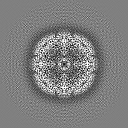
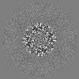
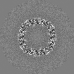
| Projections & slices | Image control
Images are generated by Spider. | ||||||||||||||||||||||||||||||||||||
| Voxel size | X=Y=Z: 0.899 Å | ||||||||||||||||||||||||||||||||||||
| Density |
| ||||||||||||||||||||||||||||||||||||
| Symmetry | Space group: 1 | ||||||||||||||||||||||||||||||||||||
| Details | EMDB XML:
|
 Z (Sec.)
 Y (Row.)
 X (Col.)


-Supplemental data
- Sample components
-Entire : Apoferritin
| Entire | Name: Apoferritin |
|---|---|
| Components |
|
-Supramolecule #1: Apoferritin
| Supramolecule | Name: Apoferritin / type: complex / ID: 1 / Parent: 0 |
|---|---|
| Source (natural) | Organism: |
| Recombinant expression | Organism:  |
| Molecular weight | Theoretical: 440 kDa/nm |
-Experimental details
-Structure determination
| Method | cryo EM |
|---|---|
 Processing Processing | single particle reconstruction |
| Aggregation state | particle |
-Sample preparation
| Concentration | 2.05 mg/mL |
|---|---|
| Buffer | pH: 7.4 / Details: PBS |
| Grid | Model: Quantifoil R2/1 / Material: COPPER/RHODIUM / Mesh: 300 / Support film - Material: CARBON / Pretreatment - Type: GLOW DISCHARGE / Details: 30 mA |
| Vitrification | Cryogen name: ETHANE / Chamber humidity: 100 % / Chamber temperature: 293 K / Instrument: FEI VITROBOT MARK IV / Details: Under anaerobic conditions. |
| Details | From Sigma (A3641) |
- Electron microscopy
| Microscope | TFS GLACIOS |
|---|---|
| Image recording | Film or detector model: GATAN K2 SUMMIT (4k x 4k) / Detector mode: COUNTING / Digitization - Sampling interval: 5.0 µm / Digitization - Frames/image: 2-60 / Number grids imaged: 1 / Number real images: 961 / Average exposure time: 5.5 sec. / Average electron dose: 1.0 e/Å2 |
| Electron beam | Acceleration voltage: 200 kV / Electron source:  FIELD EMISSION GUN FIELD EMISSION GUN |
| Electron optics | Calibrated defocus max: 2.5500000000000003 µm / Calibrated defocus min: 0.9 µm / Illumination mode: FLOOD BEAM / Imaging mode: BRIGHT FIELD / Cs: 2.7 mm / Nominal defocus max: 2.5500000000000003 µm / Nominal defocus min: 0.9 µm / Nominal magnification: 45000 |
| Sample stage | Specimen holder model: FEI TITAN KRIOS AUTOGRID HOLDER / Cooling holder cryogen: NITROGEN |
+Image processing
-Atomic model buiding 1
| Refinement | Protocol: FLEXIBLE FIT / Overall B value: 118 |
|---|
