 Movie
Movie Controller

[English] 日本語
 Yorodumi
- PDB-7plm: CryoEM reconstruction of pyruvate ferredoxin oxidoreductase (PFOR... -
+ Open data

- Basic information
| Entry | Database: PDB / ID: 7plm | |||||||||||||||
|---|---|---|---|---|---|---|---|---|---|---|---|---|---|---|---|---|
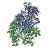
| Title | CryoEM reconstruction of pyruvate ferredoxin oxidoreductase (PFOR) in anaerobic conditions | |||||||||||||||
 Components Components | Pyruvate:ferredoxin oxidoreductase | |||||||||||||||
 Keywords Keywords | OXIDOREDUCTASE / 4Fe-4S / anaerobic / PYRUVATE CATABOLISM / Electron Transport | |||||||||||||||
| Function / homology |  Function and homology information Function and homology informationpyruvate synthase (ferredoxin) / pyruvate synthase activity / thiamine pyrophosphate binding / electron transport chain / 4 iron, 4 sulfur cluster binding / response to oxidative stress / iron ion binding / cytoplasm Similarity search - Function | |||||||||||||||
| Biological species |  Desulfocurvibacter africanus (bacteria) Desulfocurvibacter africanus (bacteria) | |||||||||||||||
| Method | ELECTRON MICROSCOPY / single particle reconstruction / cryo EM / Resolution: 2.9 Å | |||||||||||||||
 Authors Authors | Cherrier, M.V. / Vernede, X. / Fenel, D. / Martin, L. / Arragain, B. / Neumann, E. / Fontecilla Camps, J.C. / Schoehn, G. / Nicolet, Y. | |||||||||||||||
| Funding support |  France, 4items France, 4items
| |||||||||||||||
 Citation Citation | Journal: Biomolecules / Year: 2022 Title: Oxygen-Sensitive Metalloprotein Structure Determination by Cryo-Electron Microscopy. Authors: Mickaël V Cherrier / Xavier Vernède / Daphna Fenel / Lydie Martin / Benoit Arragain / Emmanuelle Neumann / Juan C Fontecilla-Camps / Guy Schoehn / Yvain Nicolet / Abstract: Metalloproteins are involved in key cell processes such as photosynthesis, respiration, and oxygen transport. However, the presence of transition metals (notably iron as a component of [Fe-S] ...Metalloproteins are involved in key cell processes such as photosynthesis, respiration, and oxygen transport. However, the presence of transition metals (notably iron as a component of [Fe-S] clusters) often makes these proteins sensitive to oxygen-induced degradation. Consequently, their study usually requires strict anaerobic conditions. Although X-ray crystallography has been the method of choice for solving macromolecular structures for many years, recently electron microscopy has also become an increasingly powerful structure-solving technique. We have used our previous experience with cryo-crystallography to develop a method to prepare cryo-EM grids in an anaerobic chamber and have applied it to solve the structures of apoferritin and the 3 [FeS]-containing pyruvate ferredoxin oxidoreductase (PFOR) at 2.40 Å and 2.90 Å resolution, respectively. The maps are of similar quality to the ones obtained under air, thereby validating our method as an improvement in the structural investigation of oxygen-sensitive metalloproteins by cryo-EM. | |||||||||||||||
| History |
|
- Structure visualization
| Structure viewer | Molecule: MolmilJmol/JSmol |
|---|
- Downloads & links
-Download
| PDBx/mmCIF format | 7plm.cif.gz | 462 KB | Display | PDBx/mmCIF format |
|---|---|---|---|---|
| PDB format | pdb7plm.ent.gz | 366.5 KB | Display | PDB format |
| PDBx/mmJSON format | 7plm.json.gz | Tree view | PDBx/mmJSON format | |
| Others |  Other downloads Other downloads |
-Validation report
| Arichive directory | https://data.pdbj.org/pub/pdb/validation_reports/pl/7plmftp://data.pdbj.org/pub/pdb/validation_reports/pl/7plm | HTTPS FTP |
|---|
-Related structure data
| Related structure data |  13493MC M: map data used to model this data C: citing same article ( |
|---|---|
| Similar structure data |

-Links
 PDBj
PDBj


- Assembly
| Deposited unit | 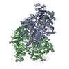
|
|---|---|
| 1 |
|
-Components
-Protein , 1 types, 2 molecules AB
| #1: Protein | Mass: 133835.109 Da / Num. of mol.: 2 Source method: isolated from a genetically manipulated source Source: (gene. exp.) Desulfocurvibacter africanus (bacteria)Gene: por / Production host: Desulfocurvibacter africanus (bacteria) / References: UniProt: P94692, pyruvate synthase (ferredoxin) |
|---|
-Non-polymers , 5 types, 126 molecules 


| #2: Chemical | ChemComp-SF4 /  Mass: 351.640 Da / Num. of mol.: 6 / Source method: obtained synthetically / Formula: Fe4S4 Mass: 351.640 Da / Num. of mol.: 6 / Source method: obtained synthetically / Formula: Fe4S4#3: Chemical |  Mass: 425.314 Da / Num. of mol.: 2 / Source method: obtained synthetically / Formula: C12H19N4O7P2S / Feature type: SUBJECT OF INVESTIGATION Mass: 425.314 Da / Num. of mol.: 2 / Source method: obtained synthetically / Formula: C12H19N4O7P2S / Feature type: SUBJECT OF INVESTIGATION#4: Chemical |  Mass: 40.078 Da / Num. of mol.: 2 / Source method: obtained synthetically / Formula: Ca Mass: 40.078 Da / Num. of mol.: 2 / Source method: obtained synthetically / Formula: Ca#5: Chemical |  Mass: 24.305 Da / Num. of mol.: 2 / Source method: obtained synthetically / Formula: Mg Mass: 24.305 Da / Num. of mol.: 2 / Source method: obtained synthetically / Formula: Mg#6: Water | ChemComp-HOH / | Mass: 18.015 Da / Num. of mol.: 114 / Source method: isolated from a natural source / Formula: H2O |
|---|
-Details
| Has ligand of interest | Y |
|---|---|
| Has protein modification | Y |
-Experimental details
-Experiment
| Experiment | Method: ELECTRON MICROSCOPY |
|---|---|
| EM experiment | Aggregation state: PARTICLE / 3D reconstruction method: single particle reconstruction |
- Sample preparation
| Component | Name: PYRUVATE FERREDOXIN OXIDOREDUCTASE / Type: COMPLEX / Entity ID: #1 / Source: NATURAL |
|---|---|
| Molecular weight | Value: 0.267360 MDa / Experimental value: NO |
| Source (natural) | Organism: Desulfocurvibacter africanus (bacteria) |
| Buffer solution | pH: 8.4 |
| Buffer component | Conc.: 10 mM / Name: Tris |
| Specimen | Conc.: 2.3 mg/ml / Embedding applied: NO / Shadowing applied: NO / Staining applied: NO / Vitrification applied: YES / Details: Monodisperse |
| Specimen support | Grid material: COPPER/RHODIUM / Grid mesh size: 300 divisions/in. / Grid type: Quantifoil R2/1 |
| Vitrification | Instrument: FEI VITROBOT MARK IV / Cryogen name: PROPANE / Humidity: 100 % / Chamber temperature: 295 K |
- Electron microscopy imaging
| Microscopy | Model: TFS GLACIOS |
|---|---|
| Electron gun | Electron source:  FIELD EMISSION GUN / Accelerating voltage: 200 kV / Illumination mode: FLOOD BEAM FIELD EMISSION GUN / Accelerating voltage: 200 kV / Illumination mode: FLOOD BEAM |
| Electron lens | Mode: BRIGHT FIELD / Nominal defocus max: 3500 nm / Nominal defocus min: 500 nm |
| Image recording | Electron dose: 60 e/Å2 / Film or detector model: GATAN K2 SUMMIT (4k x 4k) / Num. of grids imaged: 1 / Num. of real images: 1304 |
| Image scans | Movie frames/image: 60 / Used frames/image: 2-60 |
- Processing
| Software | Name: PHENIX / Version: (1.20.1_4487:phenix.real_space_refine) / Classification: refinement | ||||||||||||||||||||||||||||||||
|---|---|---|---|---|---|---|---|---|---|---|---|---|---|---|---|---|---|---|---|---|---|---|---|---|---|---|---|---|---|---|---|---|---|
| EM software |
| ||||||||||||||||||||||||||||||||
| CTF correction | Type: PHASE FLIPPING AND AMPLITUDE CORRECTION | ||||||||||||||||||||||||||||||||
| Symmetry | Point symmetry: C2 (2 fold cyclic) | ||||||||||||||||||||||||||||||||
| 3D reconstruction | Resolution: 2.9 Å / Resolution method: FSC 0.143 CUT-OFF / Num. of particles: 180868 / Symmetry type: POINT | ||||||||||||||||||||||||||||||||
| Atomic model building | Protocol: RIGID BODY FIT / Space: RECIPROCAL | ||||||||||||||||||||||||||||||||
| Atomic model building | PDB-ID: 1B0P Pdb chain-ID: A / Accession code: 1B0P / Source name: PDB / Type: experimental model | ||||||||||||||||||||||||||||||||
| Refinement | Cross valid method: THROUGHOUT | ||||||||||||||||||||||||||||||||
| Displacement parameters | Biso max: 100.48 Å2 / Biso mean: 32.3636 Å2 / Biso min: 0 Å2 |
