ムービー
ムービー コントローラー


+ データを開く

- 基本情報
基本情報
| 登録情報 | データベース: EMDB / ID: EMD-13324 | |||||||||
|---|---|---|---|---|---|---|---|---|---|---|
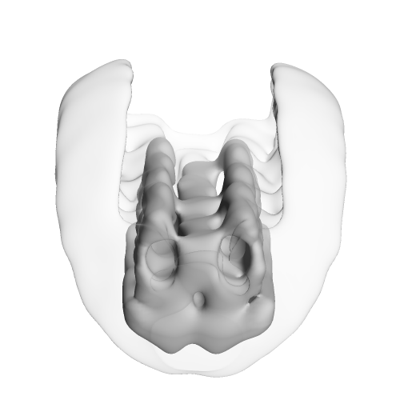
| タイトル | Central row of the protein scaffold at rod outer segment disk rims in ABCA4 knockout mice (Volta phase plate data). | |||||||||
 マップデータ マップデータ | Protein scaffold located at the outer periphery of rod outer segment disk rims in ABCA4 ko mice. The alignment mask focused on 4 repeats along the central row. Used here was a phase plate dataset. | |||||||||
 試料 試料 |
| |||||||||
| 生物種 |  | |||||||||
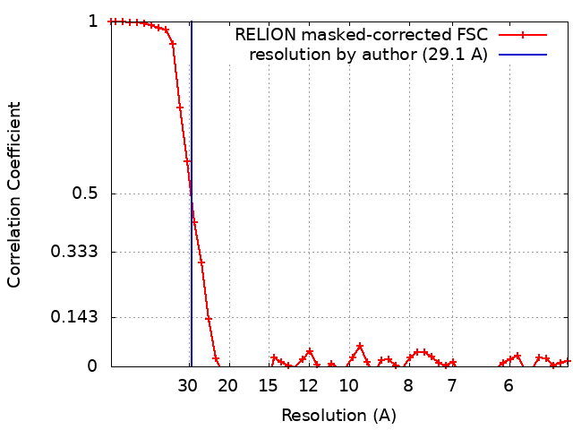
| 手法 | サブトモグラム平均法 / クライオ電子顕微鏡法 / 解像度: 29.1 Å | |||||||||
 データ登録者 データ登録者 | Poege M / Mahamid J / Imanishi SS / Plitzko JM / Palczewski K / Baumeister W | |||||||||
| 資金援助 | 1件
| |||||||||
 引用 引用 | ジャーナル: Elife / 年: 2021 タイトル: Determinants shaping the nanoscale architecture of the mouse rod outer segment. 著者: Matthias Pöge / Julia Mahamid / Sanae S Imanishi / Jürgen M Plitzko / Krzysztof Palczewski / Wolfgang Baumeister /   要旨: The unique membrane organization of the rod outer segment (ROS), the specialized sensory cilium of rod photoreceptor cells, provides the foundation for phototransduction, the initial step in vision. ...The unique membrane organization of the rod outer segment (ROS), the specialized sensory cilium of rod photoreceptor cells, provides the foundation for phototransduction, the initial step in vision. ROS architecture is characterized by a stack of identically shaped and tightly packed membrane disks loaded with the visual receptor rhodopsin. A wide range of genetic aberrations have been reported to compromise ROS ultrastructure, impairing photoreceptor viability and function. Yet, the structural basis giving rise to the remarkably precise arrangement of ROS membrane stacks and the molecular mechanisms underlying genetically inherited diseases remain elusive. Here, cryo-electron tomography (cryo-ET) performed on native ROS at molecular resolution provides insights into key structural determinants of ROS membrane architecture. Our data confirm the existence of two previously observed molecular connectors/spacers which likely contribute to the nanometer-scale precise stacking of the ROS disks. We further provide evidence that the extreme radius of curvature at the disk rims is enforced by a continuous supramolecular assembly composed of peripherin-2 (PRPH2) and rod outer segment membrane protein 1 (ROM1) oligomers. We suggest that together these molecular assemblies constitute the structural basis of the highly specialized ROS functional architecture. Our Cryo-ET data provide novel quantitative and structural information on the molecular architecture in ROS and substantiate previous results on proposed mechanisms underlying pathologies of certain PRPH2 mutations leading to blindness. | |||||||||
| 履歴 |
|
- 構造の表示
構造の表示
| ムービー |
 ムービービューア ムービービューア |
|---|---|
| 構造ビューア | EMマップ: SurfViewMolmilJmol/JSmol |
| 添付画像 |
- ダウンロードとリンク
-EMDBアーカイブ
| マップデータ | emd_13324.map.gz | 7.2 MB | EMDBマップデータ形式 | |
|---|---|---|---|---|
| ヘッダ (付随情報) | emd-13324-v30.xmlemd-13324.xml | 17.4 KB 17.4 KB | 表示 表示 | EMDBヘッダ |
| FSC (解像度算出) | emd_13324_fsc.xml | 4.7 KB | 表示 | FSCデータファイル |
| 画像 |  emd_13324.png emd_13324.png | 42.2 KB | ||
| マスクデータ | emd_13324_msk_1.map | 8 MB | マスクマップ | |
| その他 | emd_13324_additional_1.map.gzemd_13324_half_map_1.map.gzemd_13324_half_map_2.map.gz | 6.1 MB 5.9 MB 5.9 MB | ||
| アーカイブディレクトリ |  http://ftp.pdbj.org/pub/emdb/structures/EMD-13324ftp://ftp.pdbj.org/pub/emdb/structures/EMD-13324 http://ftp.pdbj.org/pub/emdb/structures/EMD-13324ftp://ftp.pdbj.org/pub/emdb/structures/EMD-13324 | HTTPS FTP |
-検証レポート
| 文書・要旨 | emd_13324_validation.pdf.gz | 468 KB | 表示 | EMDB検証レポート |
|---|---|---|---|---|
| 文書・詳細版 | emd_13324_full_validation.pdf.gz | 467.6 KB | 表示 | |
| XML形式データ | emd_13324_validation.xml.gz | 10.9 KB | 表示 | |
| CIF形式データ | emd_13324_validation.cif.gz | 13.6 KB | 表示 | |
| アーカイブディレクトリ | https://ftp.pdbj.org/pub/emdb/validation_reports/EMD-13324ftp://ftp.pdbj.org/pub/emdb/validation_reports/EMD-13324 | HTTPS FTP |
-関連構造データ
| 関連構造データ | C: 同じ文献を引用 ( |
|---|---|
| 類似構造データ | |
| 電子顕微鏡画像生データ | EMPIAR-10771 (タイトル: Cryo-electron tomography of rod outer segments in ABCA4 knockout mice acquired with Volta phase plate in focus Data size: 3.0 Data #1: Tomograms of rod outer segments in ABCA4 knockout mice acquired with Volta phase plate [reconstructed volumes]) |


-リンク
| EMDBのページ | EMDB (EBI/PDBe) / EMDataResource |
|---|
-マップ
| ファイル | ダウンロード / ファイル: emd_13324.map.gz / 形式: CCP4 / 大きさ: 8 MB / タイプ: IMAGE STORED AS FLOATING POINT NUMBER (4 BYTES) | ||||||||||||||||||||||||||||||||||||||||||||||||||||||||||||
|---|---|---|---|---|---|---|---|---|---|---|---|---|---|---|---|---|---|---|---|---|---|---|---|---|---|---|---|---|---|---|---|---|---|---|---|---|---|---|---|---|---|---|---|---|---|---|---|---|---|---|---|---|---|---|---|---|---|---|---|---|---|
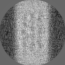
| 注釈 | Protein scaffold located at the outer periphery of rod outer segment disk rims in ABCA4 ko mice. The alignment mask focused on 4 repeats along the central row. Used here was a phase plate dataset. | ||||||||||||||||||||||||||||||||||||||||||||||||||||||||||||
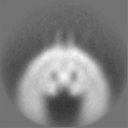
| 投影像・断面図 | 画像のコントロール
画像は Spider により作成 | ||||||||||||||||||||||||||||||||||||||||||||||||||||||||||||
| ボクセルのサイズ | X=Y=Z: 2.62 Å | ||||||||||||||||||||||||||||||||||||||||||||||||||||||||||||
| 密度 |
| ||||||||||||||||||||||||||||||||||||||||||||||||||||||||||||
| 対称性 | 空間群: 1 | ||||||||||||||||||||||||||||||||||||||||||||||||||||||||||||
| 詳細 | EMDB XML:
CCP4マップ ヘッダ情報:
| ||||||||||||||||||||||||||||||||||||||||||||||||||||||||||||
 Z (Sec.)
 Y (Row.)
 X (Col.)


-添付データ
-マスク #1
| ファイル | emd_13324_msk_1.map | ||||||||||||
|---|---|---|---|---|---|---|---|---|---|---|---|---|---|
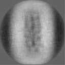
| 投影像・断面図 |
| ||||||||||||
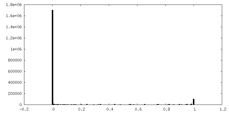
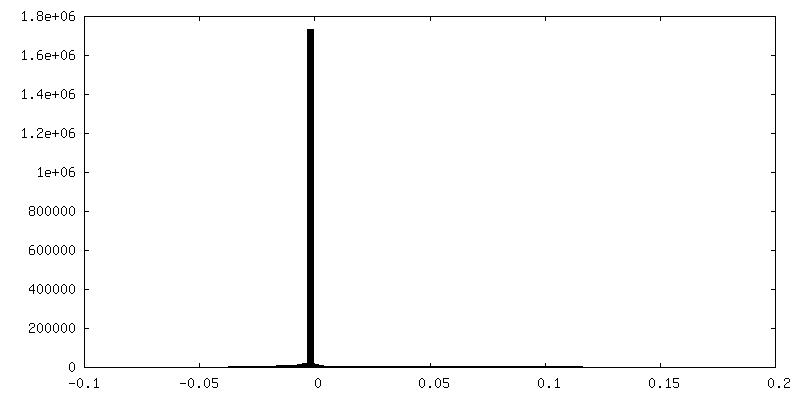
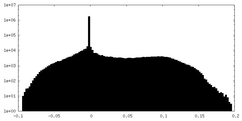
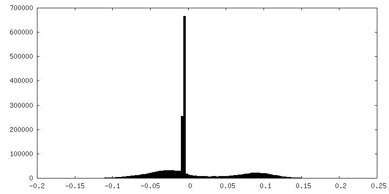
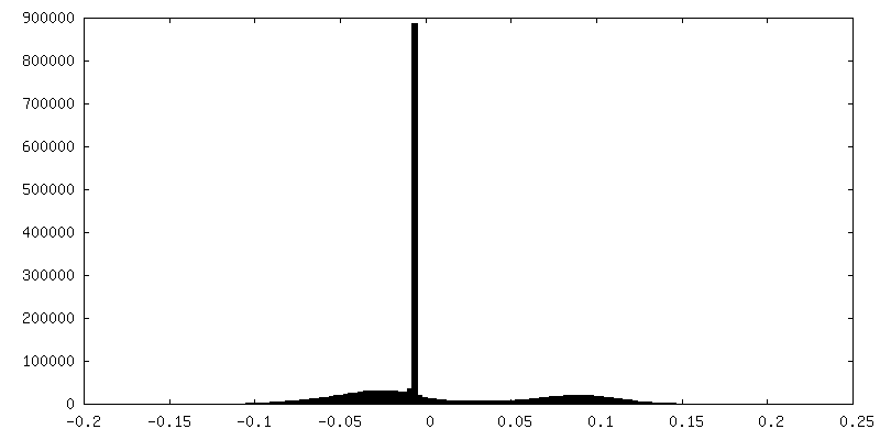
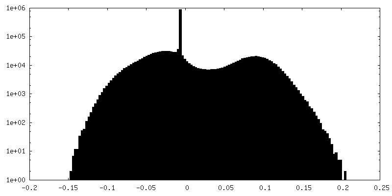
| 密度ヒストグラム |


-追加マップ: Masked volume focusing on 4 repeats along the...
| ファイル | emd_13324_additional_1.map | ||||||||||||
|---|---|---|---|---|---|---|---|---|---|---|---|---|---|
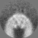
| 注釈 | Masked volume focusing on 4 repeats along the central row with its connections to the peripheral rows. | ||||||||||||
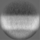
| 投影像・断面図 |
| ||||||||||||
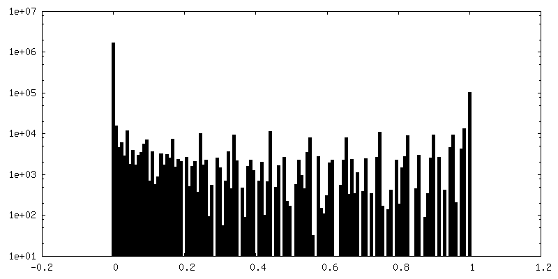
| 密度ヒストグラム |


-ハーフマップ: Unfiltered half map two as output of RELION Refine3D.
| ファイル | emd_13324_half_map_1.map | ||||||||||||
|---|---|---|---|---|---|---|---|---|---|---|---|---|---|
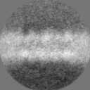
| 注釈 | Unfiltered half map two as output of RELION Refine3D. | ||||||||||||
| 投影像・断面図 |
| ||||||||||||
| 密度ヒストグラム |

-ハーフマップ: Unfiltered half map one as output of RELION Refine3D.
| ファイル | emd_13324_half_map_2.map | ||||||||||||
|---|---|---|---|---|---|---|---|---|---|---|---|---|---|
| 注釈 | Unfiltered half map one as output of RELION Refine3D. | ||||||||||||
| 投影像・断面図 |
| ||||||||||||
| 密度ヒストグラム |
- 試料の構成要素
-全体 : Rod outer segment disk rim scaffold
| 全体 | 名称: Rod outer segment disk rim scaffold |
|---|---|
| 要素 |
|
-超分子 #1: Rod outer segment disk rim scaffold
| 超分子 | 名称: Rod outer segment disk rim scaffold / タイプ: complex / ID: 1 / 親要素: 0 詳細: Protein scaffold located at the outer periphery of rod outer segment disk rims in ABCA4 ko mice - the central row. |
|---|---|
| 由来(天然) | 生物種: |
-実験情報
-構造解析
| 手法 | クライオ電子顕微鏡法 |
|---|---|
 解析 解析 | サブトモグラム平均法 |
| 試料の集合状態 | cell |
-試料調製
| 緩衝液 | pH: 7.4 / 詳細: Ringers buffer |
|---|---|
| グリッド | モデル: Quantifoil R2/1 / 材質: COPPER / メッシュ: 200 / 支持フィルム - 材質: CARBON / 支持フィルム - トポロジー: HOLEY / 前処理 - タイプ: GLOW DISCHARGE |
| 凍結 | 凍結剤: ETHANE-PROPANE / チャンバー内湿度: 90 % / チャンバー内温度: 310 K / 装置: FEI VITROBOT MARK IV |
| 詳細 | Rod outer segments were isolated by retinal detachment with a single physical disruption, applied to EM-grids and plunge frozen. |
- 電子顕微鏡法
| 顕微鏡 | FEI TITAN KRIOS |
|---|---|
| 特殊光学系 | 位相板: VOLTA PHASE PLATE / 詳細: Data was acquired in focus. |
| 撮影 | フィルム・検出器のモデル: GATAN K2 SUMMIT (4k x 4k) 検出モード: COUNTING / 平均電子線量: 1.8 e/Å2 詳細: The total accumulated dose per tilt series was ~ 100e-/A^2. |
| 電子線 | 加速電圧: 300 kV / 電子線源:  FIELD EMISSION GUN FIELD EMISSION GUN |
| 電子光学系 | 照射モード: FLOOD BEAM / 撮影モード: BRIGHT FIELD |
| 実験機器 |  モデル: Titan Krios / 画像提供: FEI Company |