 Movie
Movie Controller

[English] 日本語
 Yorodumi
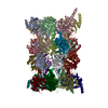
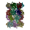
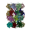
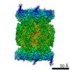
- EMDB-13183: Subtomogram average of T20S proteasome from Thermoplasma acidophilum -
+ Open data

- Basic information
| Entry | Database: EMDB / ID: EMD-13183 | |||||||||
|---|---|---|---|---|---|---|---|---|---|---|
| Title | Subtomogram average of T20S proteasome from Thermoplasma acidophilum | |||||||||
 Map data Map data | Subtomogram average of T20S proteasome from Thermoplasma acidophilum | |||||||||
 Sample Sample |
| |||||||||
| Biological species |  | |||||||||
| Method | subtomogram averaging / cryo EM / Resolution: 7.1 Å | |||||||||
 Authors Authors | Fernandez JJ / Li S | |||||||||
 Citation Citation | Journal: J Struct Biol / Year: 2021 Title: TomoAlign: A novel approach to correcting sample motion and 3D CTF in CryoET. Authors: Jose-Jesus Fernandez / Sam Li /   Abstract: TomoAlign is a software package that integrates tools to mitigate two important resolution limiting factors in cryoET, namely the beam-induced sample motion and the contrast transfer function (CTF) ...TomoAlign is a software package that integrates tools to mitigate two important resolution limiting factors in cryoET, namely the beam-induced sample motion and the contrast transfer function (CTF) of the microscope. The package is especially focused on cryoET of thick specimens where fiducial markers are required for accurate tilt-series alignment and sample motion estimation. TomoAlign models the beam-induced sample motion undergone during the tilt-series acquisition. The motion models are used to produce motion-corrected subtilt-series centered on the particles of interest. In addition, the defocus of each particle at each tilt image is determined and can be corrected, resulting in motion-corrected and CTF-corrected subtilt-series from which the subtomograms can be computed. Alternatively, the CTF information can be passed on so that CTF correction can be carried out entirely within external packages like Relion. TomoAlign serves as a versatile tool that can streamline the cryoET workflow from initial alignment of tilt-series to final subtomogram averaging during in situ structure determination. | |||||||||
| History |
|
- Structure visualization
| Movie |
 Movie viewer Movie viewer |
|---|---|
| Structure viewer | EM map: SurfViewMolmilJmol/JSmol |
| Supplemental images |
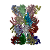
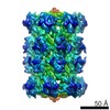
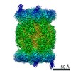
 UCSF Chimera
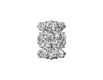
- Downloads & links
-EMDB archive
| Map data | emd_13183.map.gz | 6.3 MB | EMDB map data format | |
|---|---|---|---|---|
| Header (meta data) | emd-13183-v30.xmlemd-13183.xml | 7.1 KB 7.1 KB | Display Display | EMDB header |
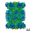
| Images |  emd_13183.png emd_13183.png | 33 KB | ||
| Archive directory |  http://ftp.pdbj.org/pub/emdb/structures/EMD-13183ftp://ftp.pdbj.org/pub/emdb/structures/EMD-13183 http://ftp.pdbj.org/pub/emdb/structures/EMD-13183ftp://ftp.pdbj.org/pub/emdb/structures/EMD-13183 | HTTPS FTP |
-Related structure data
| Related structure data | C: citing same article ( |
|---|---|
| Similar structure data |


-Links
| EMDB pages | EMDB (EBI/PDBe) / EMDataResource |
|---|
-Map
| File | Download / File: emd_13183.map.gz / Format: CCP4 / Size: 6.6 MB / Type: IMAGE STORED AS FLOATING POINT NUMBER (4 BYTES) | ||||||||||||||||||||||||||||||||||||||||||||||||||||||||||||||||||||
|---|---|---|---|---|---|---|---|---|---|---|---|---|---|---|---|---|---|---|---|---|---|---|---|---|---|---|---|---|---|---|---|---|---|---|---|---|---|---|---|---|---|---|---|---|---|---|---|---|---|---|---|---|---|---|---|---|---|---|---|---|---|---|---|---|---|---|---|---|---|
| Annotation | Subtomogram average of T20S proteasome from Thermoplasma acidophilum | ||||||||||||||||||||||||||||||||||||||||||||||||||||||||||||||||||||
| Projections & slices | Image control
Images are generated by Spider. | ||||||||||||||||||||||||||||||||||||||||||||||||||||||||||||||||||||
| Voxel size | X=Y=Z: 2.56 Å | ||||||||||||||||||||||||||||||||||||||||||||||||||||||||||||||||||||
| Density |
| ||||||||||||||||||||||||||||||||||||||||||||||||||||||||||||||||||||
| Symmetry | Space group: 1 | ||||||||||||||||||||||||||||||||||||||||||||||||||||||||||||||||||||
| Details | EMDB XML:
CCP4 map header:
| ||||||||||||||||||||||||||||||||||||||||||||||||||||||||||||||||||||
 Z (Sec.)
 Y (Row.)
 X (Col.)


-Supplemental data
- Sample components
-Entire : T20S proteasome
| Entire | Name: T20S proteasome |
|---|---|
| Components |
|
-Supramolecule #1: T20S proteasome
| Supramolecule | Name: T20S proteasome / type: complex / ID: 1 / Parent: 0 |
|---|---|
| Source (natural) | Organism: |
-Experimental details
-Structure determination
| Method | cryo EM |
|---|---|
 Processing Processing | subtomogram averaging |
| Aggregation state | particle |
-Sample preparation
| Buffer | pH: 7.5 |
|---|---|
| Vitrification | Cryogen name: ETHANE |
- Electron microscopy
| Microscope | FEI POLARA 300 |
|---|---|
| Image recording | Film or detector model: GATAN K2 SUMMIT (4k x 4k) / Average electron dose: 60.0 e/Å2 |
| Electron beam | Acceleration voltage: 300 kV / Electron source: OTHER |
| Electron optics | Illumination mode: OTHER / Imaging mode: OTHER |
| Experimental equipment |  Model: Tecnai Polara / Image courtesy: FEI Company |
-Image processing
| Final reconstruction | Applied symmetry - Point group: D7 (2x7 fold dihedral) / Resolution.type: BY AUTHOR / Resolution: 7.1 Å / Resolution method: FSC 0.5 CUT-OFF / Number subtomograms used: 3928 |
|---|---|
| Extraction | Number tomograms: 14 / Number images used: 3928 |
| Final angle assignment | Type: OTHER |
