 Movie
Movie Controller

[English] 日本語
 Yorodumi
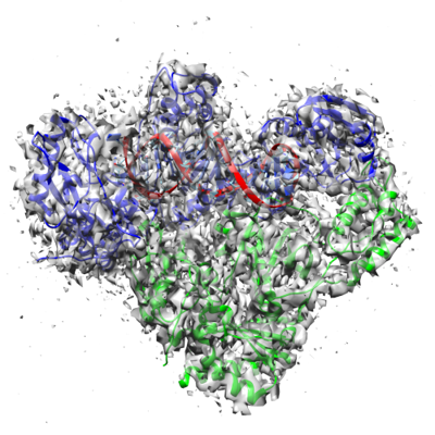
- EMDB-13156: Cryo-EM structure of HIV-1 reverse transcriptase with a DNA aptam... -
+ Open data

- Basic information
| Entry | Database: EMDB / ID: EMD-13156 | |||||||||
|---|---|---|---|---|---|---|---|---|---|---|
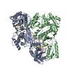
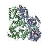
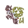
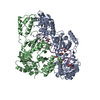
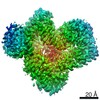
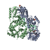
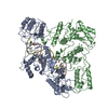
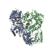
| Title | Cryo-EM structure of HIV-1 reverse transcriptase with a DNA aptamer in complex with fragment F04 at the transient P-pocket | |||||||||
 Map data Map data | ||||||||||
 Sample Sample |
| |||||||||
 Keywords Keywords | Reverse transcriptase / RT-aptamer complex / RT sliding / P-1 complex / P51 / P66 / TRANSFERASE | |||||||||
| Function / homology |  Function and homology information Function and homology informationHIV-1 retropepsin / symbiont-mediated activation of host apoptosis / retroviral ribonuclease H / exoribonuclease H / exoribonuclease H activity / DNA integration / viral genome integration into host DNA / establishment of integrated proviral latency / RNA-directed DNA polymerase / RNA stem-loop binding ...HIV-1 retropepsin / symbiont-mediated activation of host apoptosis / retroviral ribonuclease H / exoribonuclease H / exoribonuclease H activity / DNA integration / viral genome integration into host DNA / establishment of integrated proviral latency / RNA-directed DNA polymerase / RNA stem-loop binding / viral penetration into host nucleus / host multivesicular body / RNA-directed DNA polymerase activity / RNA-DNA hybrid ribonuclease activity / Transferases; Transferring phosphorus-containing groups; Nucleotidyltransferases / host cell / viral nucleocapsid / DNA recombination / DNA-directed DNA polymerase / aspartic-type endopeptidase activity / Hydrolases; Acting on ester bonds / DNA-directed DNA polymerase activity / symbiont-mediated suppression of host gene expression / viral translational frameshifting / symbiont entry into host cell / lipid binding / host cell nucleus / host cell plasma membrane / virion membrane / structural molecule activity / proteolysis / DNA binding / zinc ion binding Similarity search - Function | |||||||||
| Biological species |  Human immunodeficiency virus type 1 BH10 / synthetic construct (others) Human immunodeficiency virus type 1 BH10 / synthetic construct (others) | |||||||||
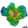
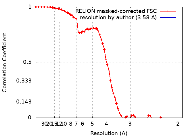
| Method | single particle reconstruction / cryo EM / Resolution: 3.58 Å | |||||||||
 Authors Authors | Singh AK / Das K | |||||||||
 Citation Citation | Journal: Nat Commun / Year: 2021 Title: Sliding of HIV-1 reverse transcriptase over DNA creates a transient P pocket - targeting P-pocket by fragment screening. Authors: Abhimanyu K Singh / Sergio E Martinez / Weijie Gu / Hoai Nguyen / Dominique Schols / Piet Herdewijn / Steven De Jonghe / Kalyan Das /  Abstract: HIV-1 reverse transcriptase (RT) slides over an RNA/DNA or dsDNA substrate while copying the viral RNA to a proviral DNA. We report a crystal structure of RT/dsDNA complex in which RT overstepped the ...HIV-1 reverse transcriptase (RT) slides over an RNA/DNA or dsDNA substrate while copying the viral RNA to a proviral DNA. We report a crystal structure of RT/dsDNA complex in which RT overstepped the primer 3'-end of a dsDNA substrate and created a transient P-pocket at the priming site. We performed a high-throughput screening of 300 drug-like fragments by X-ray crystallography that identifies two leads that bind the P-pocket, which is composed of structural elements from polymerase active site, primer grip, and template-primer that are resilient to drug-resistance mutations. Analogs of a fragment were synthesized, two of which show noticeable RT inhibition. An engineered RT/DNA aptamer complex could trap the transient P-pocket in solution, and structures of the RT/DNA complex were determined in the presence of an inhibitory fragment. A synthesized analog bound at P-pocket is further analyzed by single-particle cryo-EM. Identification of the P-pocket within HIV RT and the developed structure-based platform provide an opportunity for the design new types of polymerase inhibitors. | |||||||||
| History |
|
- Structure visualization
| Movie |
Movie viewer |
|---|---|
| Structure viewer | EM map: SurfViewMolmilJmol/JSmol |
| Supplemental images |
- Downloads & links
-EMDB archive
| Map data | emd_13156.map.gz | 6.9 MB | EMDB map data format | |
|---|---|---|---|---|
| Header (meta data) | emd-13156-v30.xmlemd-13156.xml | 22.7 KB 22.7 KB | Display Display | EMDB header |
| FSC (resolution estimation) | emd_13156_fsc.xml | 6.9 KB | Display | FSC data file |
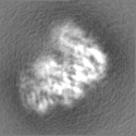
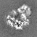
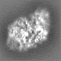
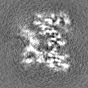
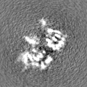
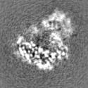
| Images |  emd_13156.png emd_13156.png | 176.6 KB | ||
| Filedesc metadata | emd-13156.cif.gz | 6.7 KB | ||
| Others | emd_13156_half_map_1.map.gzemd_13156_half_map_2.map.gz | 6.9 MB 6.9 MB | ||
| Archive directory |  http://ftp.pdbj.org/pub/emdb/structures/EMD-13156ftp://ftp.pdbj.org/pub/emdb/structures/EMD-13156 http://ftp.pdbj.org/pub/emdb/structures/EMD-13156ftp://ftp.pdbj.org/pub/emdb/structures/EMD-13156 | HTTPS FTP |
-Related structure data
| Related structure data |  7p15MC 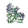 7oxqC 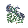 7oz2C 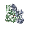 7oz5C 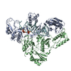 7ozwC C: citing same article ( M: atomic model generated by this map |
|---|---|
| Similar structure data |
-Links
| EMDB pages | EMDB (EBI/PDBe) / EMDataResource |
|---|---|
| Related items in Molecule of the Month |


-Map
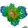
| File | Download / File: emd_13156.map.gz / Format: CCP4 / Size: 7.5 MB / Type: IMAGE STORED AS FLOATING POINT NUMBER (4 BYTES) | ||||||||||||||||||||||||||||||||||||||||||||||||||||||||||||
|---|---|---|---|---|---|---|---|---|---|---|---|---|---|---|---|---|---|---|---|---|---|---|---|---|---|---|---|---|---|---|---|---|---|---|---|---|---|---|---|---|---|---|---|---|---|---|---|---|---|---|---|---|---|---|---|---|---|---|---|---|---|
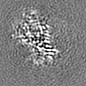
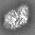
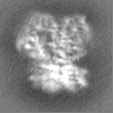
| Projections & slices | Image control
Images are generated by Spider. | ||||||||||||||||||||||||||||||||||||||||||||||||||||||||||||
| Voxel size | X=Y=Z: 0.97 Å | ||||||||||||||||||||||||||||||||||||||||||||||||||||||||||||
| Density |
| ||||||||||||||||||||||||||||||||||||||||||||||||||||||||||||
| Symmetry | Space group: 1 | ||||||||||||||||||||||||||||||||||||||||||||||||||||||||||||
| Details | EMDB XML:
CCP4 map header:
| ||||||||||||||||||||||||||||||||||||||||||||||||||||||||||||
 Z (Sec.)
 Y (Row.)
 X (Col.)


-Supplemental data
-Half map: #1
| File | emd_13156_half_map_1.map | ||||||||||||
|---|---|---|---|---|---|---|---|---|---|---|---|---|---|
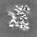
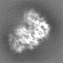
| Projections & Slices |
| ||||||||||||
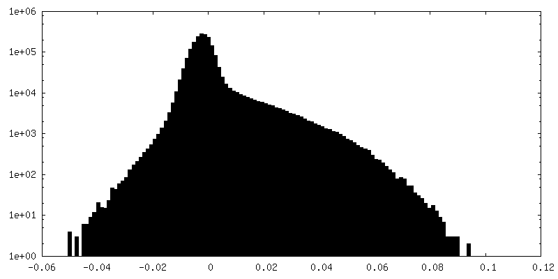
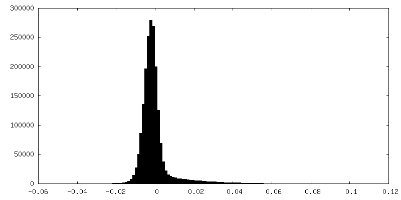
| Density Histograms |


-Half map: #2
| File | emd_13156_half_map_2.map | ||||||||||||
|---|---|---|---|---|---|---|---|---|---|---|---|---|---|
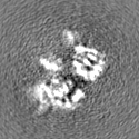
| Projections & Slices |
| ||||||||||||
| Density Histograms |

- Sample components
-Entire : Cryo-EM structure of HIV-1 reverse transcriptase with a DNA aptam...
| Entire | Name: Cryo-EM structure of HIV-1 reverse transcriptase with a DNA aptamer in complex with fragment F04 at the transient P-pocket |
|---|---|
| Components |
|
-Supramolecule #1: Cryo-EM structure of HIV-1 reverse transcriptase with a DNA aptam...
| Supramolecule | Name: Cryo-EM structure of HIV-1 reverse transcriptase with a DNA aptamer in complex with fragment F04 at the transient P-pocket type: complex / ID: 1 / Parent: 0 / Macromolecule list: #1-#3 |
|---|---|
| Source (natural) | Organism: Human immunodeficiency virus type 1 BH10 |
| Molecular weight | Theoretical: 11.43 KDa |
-Supramolecule #2: HIV-1 REVERSE TRANSCRIPTASE P66 SUBUNIT
| Supramolecule | Name: HIV-1 REVERSE TRANSCRIPTASE P66 SUBUNIT / type: complex / ID: 2 / Parent: 1 / Macromolecule list: #1 |
|---|---|
| Source (natural) | Organism: Human immunodeficiency virus type 1 BH10 |
-Supramolecule #3: HIV-1 REVERSE TRANSCRIPTASE P51 SUBUNIT
| Supramolecule | Name: HIV-1 REVERSE TRANSCRIPTASE P51 SUBUNIT / type: complex / ID: 3 / Parent: 1 / Macromolecule list: #2 |
|---|---|
| Source (natural) | Organism: Human immunodeficiency virus type 1 BH10 |
-Supramolecule #4: DNA (37-mer)
| Supramolecule | Name: DNA (37-mer) / type: complex / ID: 4 / Parent: 1 / Macromolecule list: #3 / Details: Synthetic DNA construct |
|---|---|
| Source (natural) | Organism: Human immunodeficiency virus type 1 BH10 |
-Macromolecule #1: Reverse transcriptase/ribonuclease H
| Macromolecule | Name: Reverse transcriptase/ribonuclease H / type: protein_or_peptide / ID: 1 / Number of copies: 1 / Enantiomer: LEVO / EC number: RNA-directed DNA polymerase |
|---|---|
| Source (natural) | Organism: Human immunodeficiency virus type 1 BH10 |
| Molecular weight | Theoretical: 64.047398 KDa |
| Recombinant expression | Organism:  |
| Sequence | String: MVPISPIETV PVKLKPGMDG PKVKQWPLTE EKIKALVEIC TEMEKEGKIS KIGPENPYNT PVFAIKKKDS TKWRKLVDFR ELNKRTQDF WEVQLGIPHP AGLKKKKSVT VLDVGDAYFS VPLDEDFRKY TAFTIPSINN ETPGIRYQYN VLPQGWKGSP A IFQSSMTK ...String: MVPISPIETV PVKLKPGMDG PKVKQWPLTE EKIKALVEIC TEMEKEGKIS KIGPENPYNT PVFAIKKKDS TKWRKLVDFR ELNKRTQDF WEVQLGIPHP AGLKKKKSVT VLDVGDAYFS VPLDEDFRKY TAFTIPSINN ETPGIRYQYN VLPQGWKGSP A IFQSSMTK ILEPFKKQNP DIVIYQYMDD LYVGSDLEIG QHRTKIEELR QHLLRWGLTT PDKKHQKEPP FLWMGYELHP DK WTVQPIV LPEKDSWTVN DIQKLVGKLN WASQIYPGIK VRQLSKLLRG TKALTEVIPL TEEAELELAE NREILKEPVH GVY YDPSKD LIAEIQKQGQ GQWTYQIYQE PFKNLKTGKY ARMRGAHTND VKQLTEAVQK ITTESIVIWG KTPKFKLPIQ KETW ETWWT EYWQATWIPE WEFVNTPPLV KLWYQLEKEP IVGAETFYVD GAANRETKLG KAGYVTNKGR QKVVPLTNTT NQKTE LQAI YLALQDSGLE VNIVTNSQYA LGIIQAQPDK SESELVNQII EQLIKKEKVY LAWVPAHKGI GGNEQVDKLV SA UniProtKB: Gag-Pol polyprotein |
-Macromolecule #2: Reverse transcriptase/ribonuclease H
| Macromolecule | Name: Reverse transcriptase/ribonuclease H / type: protein_or_peptide / ID: 2 / Number of copies: 1 / Enantiomer: LEVO / EC number: RNA-directed DNA polymerase |
|---|---|
| Source (natural) | Organism: Human immunodeficiency virus type 1 BH10 |
| Molecular weight | Theoretical: 50.039488 KDa |
| Recombinant expression | Organism: |
| Sequence | String: PISPIETVPV KLKPGMDGPK VKQWPLTEEK IKALVEICTE MEKEGKISKI GPENPYNTPV FAIKKKDSTK WRKLVDFREL NKRTQDFWE VQLGIPHPAG LKKKKSVTVL DVGDAYFSVP LDEDFRKYTA FTIPSINNET PGIRYQYNVL PQGWKGSPAI F QSSMTKIL ...String: PISPIETVPV KLKPGMDGPK VKQWPLTEEK IKALVEICTE MEKEGKISKI GPENPYNTPV FAIKKKDSTK WRKLVDFREL NKRTQDFWE VQLGIPHPAG LKKKKSVTVL DVGDAYFSVP LDEDFRKYTA FTIPSINNET PGIRYQYNVL PQGWKGSPAI F QSSMTKIL EPFKKQNPDI VIYQYMDDLY VGSDLEIGQH RTKIEELRQH LLRWGLTTPD KKHQKEPPFL WMGYELHPDK WT VQPIVLP EKDSWTVNDI QKLVGKLNWA SQIYPGIKVR QLSKLLRGTK ALTEVIPLTE EAELELAENR EILKEPVHGV YYD PSKDLI AEIQKQGQGQ WTYQIYQEPF KNLKTGKYAR MRGAHTNDVK QLTEAVQKIT TESIVIWGKT PKFKLPIQKE TWET WWTEY WQATWIPEWE FVNTPPLVKL WYQ UniProtKB: Gag-Pol polyprotein |
-Macromolecule #3: DNA (37-MER)
| Macromolecule | Name: DNA (37-MER) / type: dna / ID: 3 / Number of copies: 1 / Classification: DNA |
|---|---|
| Source (natural) | Organism: synthetic construct (others) |
| Molecular weight | Theoretical: 11.434332 KDa |
| Sequence | String: (DT)(DA)(DA)(DT)(DA)(DT)(OMC)(DC)(OMC)(DC) (DC)(DC)(DT)(DT)(DC)(DG)(DG)(DT)(DG) (DC)(DT)(DT)(DT)(DG)(DC)(DA)(DC)(DC)(DG) (DA)(DA)(DG)(DG)(DG)(DG)(DG)(DG) |
-Macromolecule #4: (1~{R},2~{R})-~{N}-(1~{H}-pyrazol-4-yl)-2-pyridin-3-yl-cyclopropa...
| Macromolecule | Name: (1~{R},2~{R})-~{N}-(1~{H}-pyrazol-4-yl)-2-pyridin-3-yl-cyclopropane-1-carboxamide type: ligand / ID: 4 / Number of copies: 1 / Formula: 4OI |
|---|---|
| Molecular weight | Theoretical: 228.25 Da |
| Chemical component information | 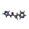 ChemComp-4OI: |
-Experimental details
-Structure determination
| Method | cryo EM |
|---|---|
 Processing Processing | single particle reconstruction |
| Aggregation state | particle |
-Sample preparation
| Concentration | 0.4 mg/mL | |||||||||
|---|---|---|---|---|---|---|---|---|---|---|
| Buffer | pH: 8 Component:
| |||||||||
| Grid | Model: Quantifoil R1.2/1.3 / Material: COPPER / Mesh: 300 / Pretreatment - Type: GLOW DISCHARGE / Pretreatment - Time: 60 sec. / Pretreatment - Atmosphere: AIR / Pretreatment - Pressure: 0.03 kPa | |||||||||
| Vitrification | Cryogen name: ETHANE / Chamber humidity: 95 % / Chamber temperature: 281 K / Instrument: LEICA EM GP |
- Electron microscopy
| Microscope | TFS GLACIOS |
|---|---|
| Image recording | Film or detector model: FEI FALCON III (4k x 4k) / Detector mode: COUNTING / Number grids imaged: 1 / Number real images: 767 / Average exposure time: 55.0 sec. / Average electron dose: 50.0 e/Å2 |
| Electron beam | Acceleration voltage: 200 kV / Electron source:  FIELD EMISSION GUN FIELD EMISSION GUN |
| Electron optics | C2 aperture diameter: 50.0 µm / Illumination mode: FLOOD BEAM / Imaging mode: BRIGHT FIELD / Cs: 2.7 mm / Nominal defocus max: 2.2 µm / Nominal defocus min: 0.8 µm / Nominal magnification: 150000 |
| Sample stage | Specimen holder model: FEI TITAN KRIOS AUTOGRID HOLDER / Cooling holder cryogen: NITROGEN |
+Image processing
-Atomic model buiding 1
| Initial model | PDB ID: Chain - Source name: PDB / Chain - Initial model type: experimental model |
|---|---|
| Refinement | Space: REAL / Protocol: FLEXIBLE FIT / Overall B value: 148.9 / Target criteria: Correlation coefficient |
| Output model | PDB-7p15: |
