 ムービー
ムービー コントローラー


+ データを開く

- 基本情報
基本情報
| 登録情報 | データベース: EMDB / ID: EMD-13152 | |||||||||
|---|---|---|---|---|---|---|---|---|---|---|
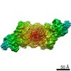
| タイトル | Negative stain EM 3D reconstruction of the Dam1 / DASH complex. | |||||||||
 マップデータ マップデータ | Dimer of the Dam1 / DASH complex. | |||||||||
 試料 試料 |
| |||||||||
| 機能・相同性 |  機能・相同性情報 機能・相同性情報protein localization to microtubule plus-end / mitotic spindle orientation checkpoint signaling / mitotic spindle polar microtubule / 2-micrometer plasmid partitioning / centromere clustering / DASH complex / protein transport along microtubule to mitotic spindle pole body / mitotic sister chromatid biorientation / mitotic spindle midzone / mitotic spindle pole body ...protein localization to microtubule plus-end / mitotic spindle orientation checkpoint signaling / mitotic spindle polar microtubule / 2-micrometer plasmid partitioning / centromere clustering / DASH complex / protein transport along microtubule to mitotic spindle pole body / mitotic sister chromatid biorientation / mitotic spindle midzone / mitotic spindle pole body / positive regulation of attachment of spindle microtubules to kinetochore / attachment of spindle microtubules to kinetochore / protein localization to microtubule / nuclear migration along microtubule / microtubule plus-end / protein localization to chromosome, centromeric region / negative regulation of microtubule depolymerization / microtubule nucleation / microtubule plus-end binding / microtubule depolymerization / mitotic sister chromatid cohesion / spindle midzone / microtubule organizing center / regulation of microtubule polymerization or depolymerization / positive regulation of microtubule polymerization / cytoplasmic microtubule / spindle assembly / spindle microtubule / spindle pole / mitotic spindle / microtubule binding / microtubule / cell division / identical protein binding 類似検索 - 分子機能 | |||||||||
| 生物種 |  | |||||||||
| 手法 | 単粒子再構成法 / ネガティブ染色法 / 解像度: 35.0 Å | |||||||||
 データ登録者 データ登録者 | Engelhard L / Bourque C / Klink BU / Gatsogiannis C | |||||||||
| 資金援助 |  ドイツ, 1件 ドイツ, 1件
| |||||||||
 引用 引用 | ジャーナル: EMBO J / 年: 2021 タイトル: Phospho-regulated Bim1/EB1 interactions trigger Dam1c ring assembly at the budding yeast outer kinetochore. 著者: Alexander Dudziak / Lena Engelhard / Cole Bourque / Björn Udo Klink / Pascaline Rombaut / Nikolay Kornakov / Karolin Jänen / Franz Herzog / Christos Gatsogiannis / Stefan Westermann / 要旨: Kinetochores form the link between chromosomes and microtubules of the mitotic spindle. The heterodecameric Dam1 complex (Dam1c) is a major component of the Saccharomyces cerevisiae outer ...Kinetochores form the link between chromosomes and microtubules of the mitotic spindle. The heterodecameric Dam1 complex (Dam1c) is a major component of the Saccharomyces cerevisiae outer kinetochore, assembling into 3 MDa-sized microtubule-embracing rings, but how ring assembly is specifically initiated in vivo remains to be understood. Here, we describe a molecular pathway that provides local control of ring assembly during the establishment of sister kinetochore bi-orientation. We show that Dam1c and the general microtubule plus end-associated protein (+TIP) Bim1/EB1 form a stable complex depending on a conserved motif in the Duo1 subunit of Dam1c. EM analyses reveal that Bim1 crosslinks protrusion domains of adjacent Dam1c heterodecamers and promotes the formation of oligomers with defined curvature. Disruption of the Dam1c-Bim1 interaction impairs kinetochore localization of Dam1c in metaphase and delays mitosis. Phosphorylation promotes Dam1c-Bim1 binding by relieving an intramolecular inhibition of the Dam1 C-terminus. In addition, Bim1 recruits Bik1/CLIP-170 to Dam1c and induces formation of full rings even in the absence of microtubules. Our data help to explain how new kinetochore end-on attachments are formed during the process of attachment error correction. | |||||||||
| 履歴 |
|
- 構造の表示
構造の表示
| ムービー |
ムービービューア |
|---|---|
| 構造ビューア | EMマップ: SurfViewMolmilJmol/JSmol |
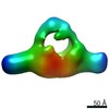
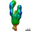
| 添付画像 |
 UCSF Chimera
- ダウンロードとリンク
-EMDBアーカイブ
| マップデータ | emd_13152.map.gz | 460 KB | EMDBマップデータ形式 | |
|---|---|---|---|---|
| ヘッダ (付随情報) | emd-13152-v30.xmlemd-13152.xml | 17.5 KB 17.5 KB | 表示 表示 | EMDBヘッダ |
| 画像 | 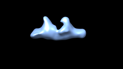 emd_13152.png emd_13152.png | 13.9 KB | ||
| アーカイブディレクトリ |  http://ftp.pdbj.org/pub/emdb/structures/EMD-13152ftp://ftp.pdbj.org/pub/emdb/structures/EMD-13152 http://ftp.pdbj.org/pub/emdb/structures/EMD-13152ftp://ftp.pdbj.org/pub/emdb/structures/EMD-13152 | HTTPS FTP |
-関連構造データ
-リンク
| EMDBのページ | EMDB (EBI/PDBe) / EMDataResource |
|---|---|
| 「今月の分子」の関連する項目 |

-マップ
| ファイル | ダウンロード / ファイル: emd_13152.map.gz / 形式: CCP4 / 大きさ: 1.7 MB / タイプ: IMAGE STORED AS FLOATING POINT NUMBER (4 BYTES) | ||||||||||||||||||||||||||||||||||||||||||||||||||||||||||||
|---|---|---|---|---|---|---|---|---|---|---|---|---|---|---|---|---|---|---|---|---|---|---|---|---|---|---|---|---|---|---|---|---|---|---|---|---|---|---|---|---|---|---|---|---|---|---|---|---|---|---|---|---|---|---|---|---|---|---|---|---|---|
| 注釈 | Dimer of the Dam1 / DASH complex. | ||||||||||||||||||||||||||||||||||||||||||||||||||||||||||||
| 投影像・断面図 | 画像のコントロール
画像は Spider により作成 | ||||||||||||||||||||||||||||||||||||||||||||||||||||||||||||
| ボクセルのサイズ | X=Y=Z: 4.92 Å | ||||||||||||||||||||||||||||||||||||||||||||||||||||||||||||
| 密度 |
| ||||||||||||||||||||||||||||||||||||||||||||||||||||||||||||
| 対称性 | 空間群: 1 | ||||||||||||||||||||||||||||||||||||||||||||||||||||||||||||
| 詳細 | EMDB XML:
CCP4マップ ヘッダ情報:
| ||||||||||||||||||||||||||||||||||||||||||||||||||||||||||||
 Z (Sec.)
 Y (Row.)
 X (Col.)


-添付データ
- 試料の構成要素
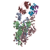
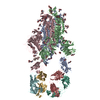
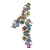
+全体 : A dimer of the Dam1 / DASH complex.
+超分子 #1: A dimer of the Dam1 / DASH complex.
+分子 #1: Ask1p of the Dam1c of S. cerevisiae
+分子 #2: Dam1p of the Dam1c of S. cerevisiae
+分子 #3: Spc34p of the Dam1c of S. cerevisiae
+分子 #4: Duo1p of the Dam1c of S. cerevisiae
+分子 #5: Spc19p of the Dam1c of S. cerevisiae
+分子 #6: Dad2p of the Dam1c of S. cerevisiae
+分子 #7: Dad1p of the Dam1c of S. cerevisiae
+分子 #8: Dad3p of the Dam1c of S. cerevisiae
+分子 #9: Dad4p of the Dam1c of S. cerevisiae
+分子 #10: Hsk3p of the Dam1c of S. cerevisiae
-実験情報
-構造解析
| 手法 | ネガティブ染色法 |
|---|---|
 解析 解析 | 単粒子再構成法 |
| 試料の集合状態 | particle |
-試料調製
| 濃度 | 0.01 mg/mL |
|---|---|
| 緩衝液 | pH: 7.4 / 構成要素: (式: HEPES, NaCl, TCEP) |
| 染色 | タイプ: NEGATIVE / 材質: Uranyl formate 詳細: 4 microliters of sample were applied onto freshly glow-discharged carbon-coated copper grids (Agar Scientific, 6400C). After an incubation of 2 minutes, the sample was blotted with Whatman no. ...詳細: 4 microliters of sample were applied onto freshly glow-discharged carbon-coated copper grids (Agar Scientific, 6400C). After an incubation of 2 minutes, the sample was blotted with Whatman no. 4 filter paper, washed 2 times with dd H20 and stained with 75% uranyl formate, blotted immediately, and then stained again and incubated for 1 minute before final blotting. The sample was then air-dried for 4 minutes. |
| グリッド | モデル: Homemade / 支持フィルム - 材質: CARBON / 前処理 - タイプ: GLOW DISCHARGE |
| 詳細 | 25 mM HEPES, pH 7.4, 200 mM NaCl, 1 mM MgCl2, and 0.5 mM TECP. |
- 電子顕微鏡法
| 顕微鏡 | JEOL 1400 |
|---|---|
| 撮影 | フィルム・検出器のモデル: TVIPS TEMCAM-F416 (4k x 4k) 平均電子線量: 5.0 e/Å2 |
| 電子線 | 加速電圧: 120 kV / 電子線源: LAB6 |
| 電子光学系 | 照射モード: SPOT SCAN / 撮影モード: BRIGHT FIELD / Cs: 3.4 mm |
| 試料ステージ | 試料ホルダーモデル: JEOL |
-画像解析
| 初期モデル | モデルのタイプ: INSILICO MODEL In silico モデル: Initial Model was computed using SPHIRE (VIPER model) |
|---|---|
| 最終 再構成 | 想定した対称性 - 点群: C1 (非対称) / 解像度のタイプ: BY AUTHOR / 解像度: 35.0 Å / 解像度の算出法: OTHER / 使用した粒子像数: 28140 |
| 初期 角度割当 | タイプ: NOT APPLICABLE |
| 最終 角度割当 | タイプ: NOT APPLICABLE |
