Journal: EMBO J / Year: 2021 Title: Phospho-regulated Bim1/EB1 interactions trigger Dam1c ring assembly at the budding yeast outer kinetochore. Authors: Alexander Dudziak / Lena Engelhard / Cole Bourque / Björn Udo Klink / Pascaline Rombaut / Nikolay Kornakov / Karolin Jänen / Franz Herzog / Christos Gatsogiannis / Stefan Westermann / Abstract: Kinetochores form the link between chromosomes and microtubules of the mitotic spindle. The heterodecameric Dam1 complex (Dam1c) is a major component of the Saccharomyces cerevisiae outer ...Kinetochores form the link between chromosomes and microtubules of the mitotic spindle. The heterodecameric Dam1 complex (Dam1c) is a major component of the Saccharomyces cerevisiae outer kinetochore, assembling into 3 MDa-sized microtubule-embracing rings, but how ring assembly is specifically initiated in vivo remains to be understood. Here, we describe a molecular pathway that provides local control of ring assembly during the establishment of sister kinetochore bi-orientation. We show that Dam1c and the general microtubule plus end-associated protein (+TIP) Bim1/EB1 form a stable complex depending on a conserved motif in the Duo1 subunit of Dam1c. EM analyses reveal that Bim1 crosslinks protrusion domains of adjacent Dam1c heterodecamers and promotes the formation of oligomers with defined curvature. Disruption of the Dam1c-Bim1 interaction impairs kinetochore localization of Dam1c in metaphase and delays mitosis. Phosphorylation promotes Dam1c-Bim1 binding by relieving an intramolecular inhibition of the Dam1 C-terminus. In addition, Bim1 recruits Bik1/CLIP-170 to Dam1c and induces formation of full rings even in the absence of microtubules. Our data help to explain how new kinetochore end-on attachments are formed during the process of attachment error correction.
History
Deposition
Jun 30, 2021
-
Header (metadata) release
Jul 28, 2021
-
Map release
Jul 28, 2021
-
Update
Sep 29, 2021
-
Current status
Sep 29, 2021
Processing site: PDBe / Status: Released
-
Structure visualization
Movie
Surface view with section colored by density value
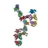
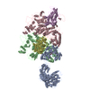
Protein or peptide: Ask1 protein of the Dam1c from S. cerevisiae
Protein or peptide: Dam1 protein of the Dam1c from S. cerevisiae
Protein or peptide: Dam1 protein of the Dam1c from S. cerevisiae
Protein or peptide: Duo1 protein of the Dam1c from S. cerevisiae
Protein or peptide: Spc19 protein of the Dam1c from S. cerevisiae
Protein or peptide: Dad2p of the Dam1c from S. cerevisiae
Protein or peptide: Dad1p of the Dam1c from S. cerevisiae
Protein or peptide: Dad3p of the Dam1c from S. cerevisiae
Protein or peptide: Dad4p of the Dam1c from S. cerevisiae
Protein or peptide: Hsk3p of the Dam1c from S. cerevisiae
Protein or peptide: Bim1p of S. cerevisiae
+
Supramolecule #1: Dam1pBim1p Complex
Supramolecule
Name: Dam1pBim1p Complex / type: complex / ID: 1 / Parent: 0 / Macromolecule list: all Details: An unknown number of copies of Bim1p (2-4) binds to a dimeric complex formed by two Dam1c heterodecamers
pH: 7.4 / Component: (Formula: HEPES, NaCl, TCEP, Glycerol) Details: The sample was crosslinked using 0.5 % glutaraldehyde. The crosslinking reaction was stopped after 60 seconds by the addition of TRIS
Staining
Type: NEGATIVE / Material: Uranyl Formate Details: 4 microl of complex were applied on freshly glow-discharged carbon-coated copper grids (Agar Scientific, G400C). After an incubation of 2 minutes, the sample was blotted with Whatman no. 4 ...Details: 4 microl of complex were applied on freshly glow-discharged carbon-coated copper grids (Agar Scientific, G400C). After an incubation of 2 minutes, the sample was blotted with Whatman no. 4 filter paper, washed 2 times with ddH2O and stained with 0.75 % uranyl formate.
Grid
Model: Homemade / Support film - Material: CARBON / Pretreatment - Type: GLOW DISCHARGE
Details
25 mM HEPES, pH 7.4, 200 mM NaCl, 1 mM MgCl2, 0.5 mM TCEP and 2.5 (v/v) % glycerol
-
Electron microscopy
Microscope
JEOL 1400
Image recording
Film or detector model: TVIPS TEMCAM-F416 (4k x 4k) / Average electron dose: 5.0 e/Å2
Electron beam
Acceleration voltage: 120 kV / Electron source: LAB6
Electron optics
Illumination mode: SPOT SCAN / Imaging mode: BRIGHT FIELD / Cs: 3.4 mm
Sample stage
Specimen holder model: JEOL
-
Image processing
Startup model
Type of model: INSILICO MODEL In silico model: Initial Model was computed using SPHIRE (VIPER module)
Final reconstruction
Applied symmetry - Point group: C1 (asymmetric) / Resolution.type: BY AUTHOR / Resolution: 35.0 Å / Resolution method: OTHER / Software - Name: SPHIRE / Number images used: 36980
Initial angle assignment
Type: NOT APPLICABLE
Final angle assignment
Type: NOT APPLICABLE
+
About Yorodumi
-
News
-
Feb 9, 2022. New format data for meta-information of EMDB entries
New format data for meta-information of EMDB entries
Version 3 of the EMDB header file is now the official format.
The previous official version 1.9 will be removed from the archive.
In the structure databanks used in Yorodumi, some data are registered as the other names, "COVID-19 virus" and "2019-nCoV". Here are the details of the virus and the list of structure data.
Jan 31, 2019. EMDB accession codes are about to change! (news from PDBe EMDB page)
EMDB accession codes are about to change! (news from PDBe EMDB page)
The allocation of 4 digits for EMDB accession codes will soon come to an end. Whilst these codes will remain in use, new EMDB accession codes will include an additional digit and will expand incrementally as the available range of codes is exhausted. The current 4-digit format prefixed with “EMD-” (i.e. EMD-XXXX) will advance to a 5-digit format (i.e. EMD-XXXXX), and so on. It is currently estimated that the 4-digit codes will be depleted around Spring 2019, at which point the 5-digit format will come into force.
The EM Navigator/Yorodumi systems omit the EMD- prefix.
Related info.:Q: What is EMD? / ID/Accession-code notation in Yorodumi/EM Navigator
Yorodumi is a browser for structure data from EMDB, PDB, SASBDB, etc.
This page is also the successor to EM Navigator detail page, and also detail information page/front-end page for Omokage search.
The word "yorodu" (or yorozu) is an old Japanese word meaning "ten thousand". "mi" (miru) is to see.
Related info.:EMDB / PDB / SASBDB / Comparison of 3 databanks / Yorodumi Search / Aug 31, 2016. New EM Navigator & Yorodumi / Yorodumi Papers / Jmol/JSmol / Function and homology information / Changes in new EM Navigator and Yorodumi
 Movie
Movie Controller


 Open data
 Basic information
 Map data
 Sample
 Function and homology information
 Authors
 Germany, 1 items
  Citation
 Structure visualization
 UCSF Chimera
 Downloads & links
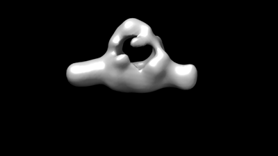 emd_13151.png
 http://ftp.pdbj.org/pub/emdb/structures/EMD-13151
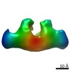


 Z (Sec.)
 Y (Row.)
 X (Col.)


 Sample components
 Processing
 Electron microscopy