: / Formation of RNA Pol II elongation complex / Formation of the Early Elongation Complex / Transcriptional regulation by small RNAs / RNA Polymerase II Pre-transcription Events / TP53 Regulates Transcription of DNA Repair Genes / FGFR2 alternative splicing / RNA polymerase II transcribes snRNA genes / mRNA Capping / mRNA Splicing - Minor Pathway ...: / Formation of RNA Pol II elongation complex / Formation of the Early Elongation Complex / Transcriptional regulation by small RNAs / RNA Polymerase II Pre-transcription Events / TP53 Regulates Transcription of DNA Repair Genes / FGFR2 alternative splicing / RNA polymerase II transcribes snRNA genes / mRNA Capping / mRNA Splicing - Minor Pathway / Processing of Capped Intron-Containing Pre-mRNA / RNA Polymerase II Promoter Escape / RNA Polymerase II Transcription Pre-Initiation And Promoter Opening / RNA Polymerase II Transcription Initiation / RNA Polymerase II Transcription Elongation / RNA Polymerase II Transcription Initiation And Promoter Clearance / RNA Pol II CTD phosphorylation and interaction with CE / Estrogen-dependent gene expression / Formation of TC-NER Pre-Incision Complex / Dual incision in TC-NER / Gap-filling DNA repair synthesis and ligation in TC-NER / mRNA Splicing - Major Pathway / organelle membrane / positive regulation of nuclear-transcribed mRNA poly(A) tail shortening / maintenance of transcriptional fidelity during transcription elongation by RNA polymerase II / positive regulation of translational initiation / RNA polymerase I complex / RNA polymerase III complex / transcription elongation by RNA polymerase I / RNA polymerase II, core complex / tRNA transcription by RNA polymerase III / transcription by RNA polymerase I / transcription-coupled nucleotide-excision repair / translation initiation factor binding / DNA-directed RNA polymerase complex / transcription initiation at RNA polymerase II promoter / P-body / ribonucleoside binding / fibrillar center / DNA-directed RNA polymerase / DNA-directed RNA polymerase activity / single-stranded DNA binding / transcription by RNA polymerase II / nucleic acid binding / protein dimerization activity / nuclear speck / single-stranded RNA binding / nucleotide binding / hydrolase activity / RNA-directed RNA polymerase activity / nucleolus / DNA-templated transcription / DNA binding / zinc ion binding / nucleoplasm / nucleus / cytosol Similarity search - Function
DNA-directed RNA polymerase subunit beta / DNA-directed RNA polymerase II subunit RPB4 / DNA-directed RNA polymerase II subunit RPB3 / DNA-directed RNA polymerase II subunit RPB11-a / DNA-directed RNA polymerases I, II, and III subunit RPABC3 / DNA-directed RNA polymerases I, II, and III subunit RPABC2 / DNA-directed RNA polymerase II subunit RPB7 / DNA-directed RNA polymerases I, II, and III subunit RPABC4 / DNA-directed RNA polymerases I, II, and III subunit RPABC1 / DNA-directed RNA polymerase II subunit RPB9 Similarity search - Component
Biological species
Sus scrofa domesticus (domestic pig)
Method
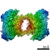
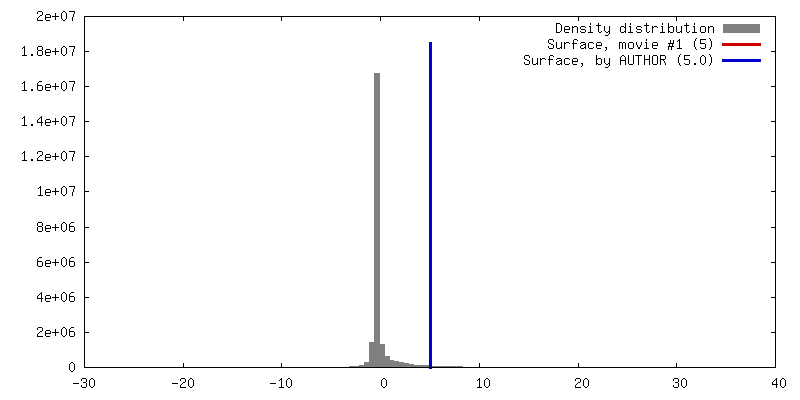
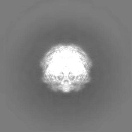
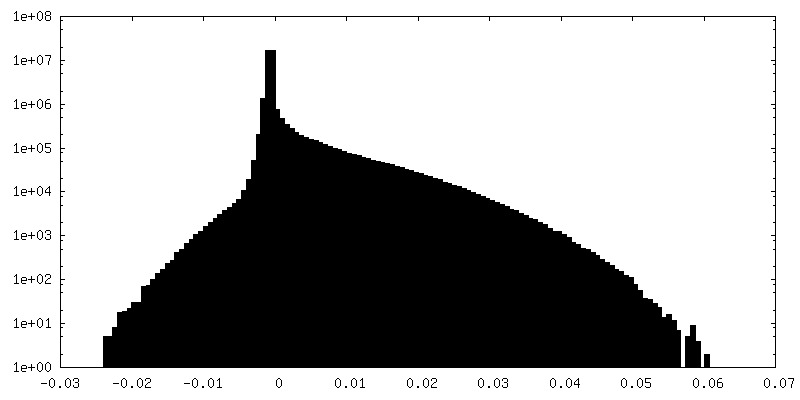
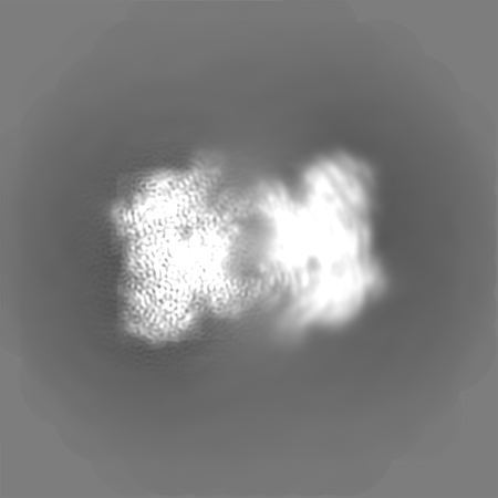
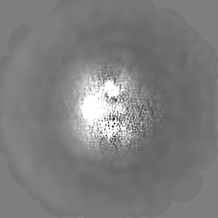
single particle reconstruction / cryo EM / Resolution: 3.5 Å
H2020 Marie Curie Actions of the European Commission
894862
Germany
German Research Foundation (DFG)
EXC 2067/1 39072994
Germany
German Research Foundation (DFG)
SFB860
Germany
German Research Foundation (DFG)
SPP2191
Germany
European Research Council (ERC)
CHROMATRANS 882357
Germany
Citation
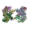
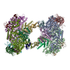
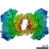
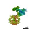
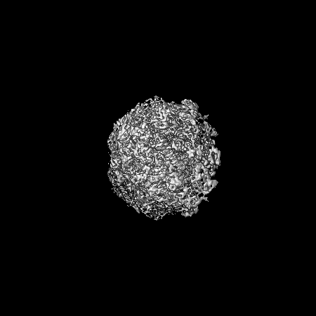
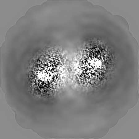
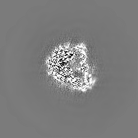
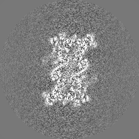
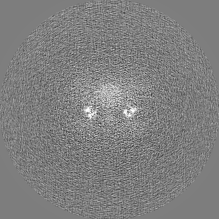
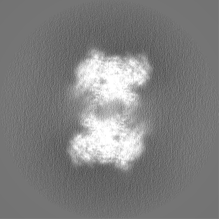
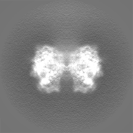
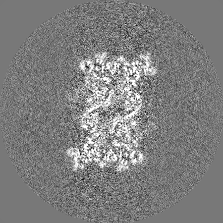
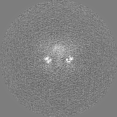
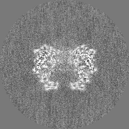
Journal: Nucleic Acids Res / Year: 2021 Title: Structure of an inactive RNA polymerase II dimer. Authors: Shintaro Aibara / Christian Dienemann / Patrick Cramer / Abstract: Eukaryotic gene transcription is carried out by three RNA polymerases: Pol I, Pol II and Pol III. Although it has long been known that Pol I can form homodimers, it is unclear whether and how the two ...Eukaryotic gene transcription is carried out by three RNA polymerases: Pol I, Pol II and Pol III. Although it has long been known that Pol I can form homodimers, it is unclear whether and how the two other RNA polymerases dimerize. Here we present the cryo-electron microscopy (cryo-EM) structure of a mammalian Pol II dimer at 3.5 Å resolution. The structure differs from the Pol I dimer and reveals that one Pol II copy uses its RPB4-RPB7 stalk to penetrate the active centre cleft of the other copy, and vice versa, giving rise to a molecular handshake. The polymerase clamp domain is displaced and mobile, and the RPB7 oligonucleotide-binding fold mimics the DNA-RNA hybrid that occupies the cleft during active transcription. The Pol II dimer is incompatible with nucleic acid binding as required for transcription and may represent an inactive storage form of the polymerase.
History
Deposition
Jun 28, 2021
-
Header (metadata) release
Oct 6, 2021
-
Map release
Oct 6, 2021
-
Update
Jul 17, 2024
-
Current status
Jul 17, 2024
Processing site: PDBe / Status: Released
-
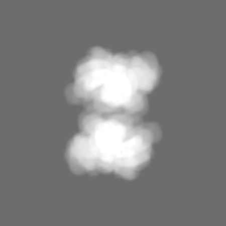
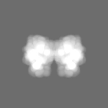
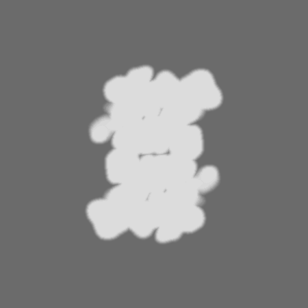
Structure visualization
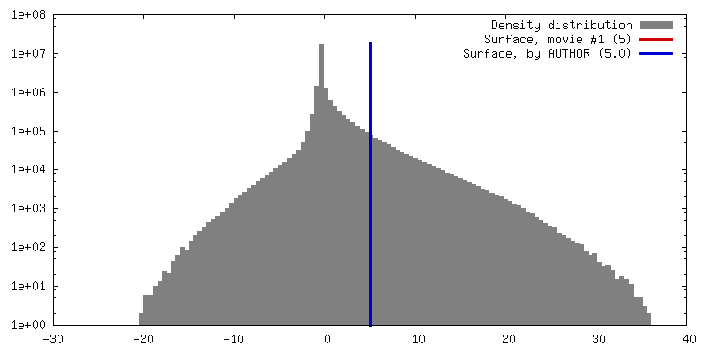
Movie
Surface view with section colored by density value
In the structure databanks used in Yorodumi, some data are registered as the other names, "COVID-19 virus" and "2019-nCoV". Here are the details of the virus and the list of structure data.
Jan 31, 2019. EMDB accession codes are about to change! (news from PDBe EMDB page)
EMDB accession codes are about to change! (news from PDBe EMDB page)
The allocation of 4 digits for EMDB accession codes will soon come to an end. Whilst these codes will remain in use, new EMDB accession codes will include an additional digit and will expand incrementally as the available range of codes is exhausted. The current 4-digit format prefixed with “EMD-” (i.e. EMD-XXXX) will advance to a 5-digit format (i.e. EMD-XXXXX), and so on. It is currently estimated that the 4-digit codes will be depleted around Spring 2019, at which point the 5-digit format will come into force.
The EM Navigator/Yorodumi systems omit the EMD- prefix.
Related info.:Q: What is EMD? / ID/Accession-code notation in Yorodumi/EM Navigator
Yorodumi is a browser for structure data from EMDB, PDB, SASBDB, etc.
This page is also the successor to EM Navigator detail page, and also detail information page/front-end page for Omokage search.
The word "yorodu" (or yorozu) is an old Japanese word meaning "ten thousand". "mi" (miru) is to see.
Related info.:EMDB / PDB / SASBDB / Comparison of 3 databanks / Yorodumi Search / Aug 31, 2016. New EM Navigator & Yorodumi / Yorodumi Papers / Jmol/JSmol / Function and homology information / Changes in new EM Navigator and Yorodumi
 Movie
Movie Controller


 Open data
 Basic information
 Map data
 Sample
 Keywords
 Function and homology information
 Authors
 Germany, 5 items
  Citation
 Structure visualization
 Downloads & links
 emd_13129.png
 http://ftp.pdbj.org/pub/emdb/structures/EMD-13129
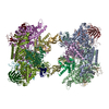


 X (Sec.)
 Y (Row.)
 Z (Col.)


 Sample components
 Processing
 Electron microscopy
 FIELD EMISSION GUN

