- EMDB-13073: The structure of MutS bound to two molecules of ADP -
+
データを開く
IDまたはキーワード:
読み込み中...
-
基本情報
登録情報
データベース: EMDB / ID: EMD-13073
タイトル
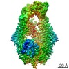
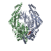
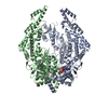
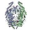
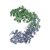
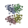
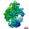
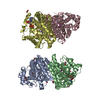
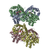
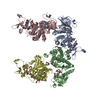
The structure of MutS bound to two molecules of ADP
マップデータ
試料
細胞器官・細胞要素: DNA mismatch repair protein MutS
タンパク質・ペプチド: DNA mismatch repair protein MutS
リガンド: ADENOSINE-5'-DIPHOSPHATE
キーワード
DNA mismatch repair protein / DNA BINDING PROTEIN
機能・相同性
機能・相同性情報
mismatched DNA binding / mismatch repair / damaged DNA binding / ATP binding 類似検索 - 分子機能
DNA mismatch repair protein MutS / DNA mismatch repair protein MutS/MSH / DNA mismatch repair protein MutS-like, N-terminal / DNA mismatch repair protein MutS, connector domain / DNA mismatch repair protein MutS, clamp / DNA mismatch repair protein MutS, N-terminal / MutS, connector domain superfamily / MutS domain I / MutS domain II / MutS family domain IV ...DNA mismatch repair protein MutS / DNA mismatch repair protein MutS/MSH / DNA mismatch repair protein MutS-like, N-terminal / DNA mismatch repair protein MutS, connector domain / DNA mismatch repair protein MutS, clamp / DNA mismatch repair protein MutS, N-terminal / MutS, connector domain superfamily / MutS domain I / MutS domain II / MutS family domain IV / MutS domain III / DNA mismatch repair protein MutS, C-terminal / DNA mismatch repair protein MutS, core / DNA mismatch repair protein MutS, core domain superfamily / MutS domain V / DNA mismatch repair proteins mutS family signature. / DNA-binding domain of DNA mismatch repair MUTS family / ATPase domain of DNA mismatch repair MUTS family / P-loop containing nucleoside triphosphate hydrolase 類似検索 - ドメイン・相同性
ジャーナル: Nat Struct Mol Biol / 年: 2022 タイトル: Cryogenic electron microscopy structures reveal how ATP and DNA binding in MutS coordinates sequential steps of DNA mismatch repair. 著者: Alessandro Borsellini / Vladislav Kunetsky / Peter Friedhoff / Meindert H Lamers / 要旨: DNA mismatch repair detects and corrects mismatches introduced during DNA replication. The protein MutS scans for mismatches and coordinates the repair cascade. During this process, MutS undergoes ...DNA mismatch repair detects and corrects mismatches introduced during DNA replication. The protein MutS scans for mismatches and coordinates the repair cascade. During this process, MutS undergoes multiple conformational changes in response to ATP binding, hydrolysis and release, but how ATP induces the various MutS conformations is incompletely understood. Here we present four cryogenic electron microscopy structures of Escherichia coli MutS at sequential stages of the ATP hydrolysis cycle that reveal how ATP binding and hydrolysis induce closing and opening of the MutS dimer, respectively. Biophysical analysis demonstrates how DNA binding modulates the ATPase cycle by prevention of hydrolysis during scanning and mismatch binding, while preventing ADP release in the sliding clamp state. Nucleotide release is achieved when MutS encounters single-stranded DNA that is produced during removal of the daughter strand. The combination of ATP binding and hydrolysis and its modulation by DNA enables MutS to adopt the different conformations needed to coordinate the sequential steps of the mismatch repair cascade.
 ムービー
ムービー コントローラー


 データを開く
 基本情報
基本情報 マップデータ
 試料
試料 キーワード
キーワード 機能・相同性情報
 データ登録者
 引用
引用

 構造の表示
構造の表示
 ダウンロードとリンク
 emd_13073.png
 http://ftp.pdbj.org/pub/emdb/structures/EMD-13073


 Z (Sec.)
 Y (Row.)
 X (Col.)


 試料の構成要素
 解析
解析 電子顕微鏡法
 FIELD EMISSION GUN