[English] 日本語
Yorodumi
- EMDB-12883: A cooperative PNPase-Hfq-RNA carrier complex facilitates bacteria... -

+
Open data


ID or keywords:

Loading...

-
Basic information

Entry
Database: EMDB / ID: EMD-12883
TitleA cooperative PNPase-Hfq-RNA carrier complex facilitates bacterial riboregulation. apo-PNPase
Map dataSharpened map, b-factor -99
Sample
  • Complex: PNPase
    • Protein or peptide: Polyribonucleotide nucleotidyltransferasePolynucleotide phosphorylase
  • Ligand: water
Function / homology
Function and homology information


polyribonucleotide nucleotidyltransferase / polyribonucleotide nucleotidyltransferase activity / bacterial degradosome / cyclic-di-GMP binding / RNA catabolic process / mRNA catabolic process / RNA processing / 3'-5'-RNA exonuclease activity / response to heat / magnesium ion binding ...polyribonucleotide nucleotidyltransferase / polyribonucleotide nucleotidyltransferase activity / bacterial degradosome / cyclic-di-GMP binding / RNA catabolic process / mRNA catabolic process / RNA processing / 3'-5'-RNA exonuclease activity / response to heat / magnesium ion binding / RNA binding / membrane / identical protein binding / cytosol
Similarity search - Function
Polyribonucleotide nucleotidyltransferase / Polyribonucleotide nucleotidyltransferase, RNA-binding domain / Polyribonucleotide nucleotidyltransferase, RNA-binding domain superfamily / Polyribonucleotide nucleotidyltransferase, RNA binding domain / Exoribonuclease, phosphorolytic domain 2 / 3' exoribonuclease family, domain 2 / Exoribonuclease, phosphorolytic domain 1 / PNPase/RNase PH domain superfamily / Exoribonuclease, PH domain 2 superfamily / 3' exoribonuclease family, domain 1 ...Polyribonucleotide nucleotidyltransferase / Polyribonucleotide nucleotidyltransferase, RNA-binding domain / Polyribonucleotide nucleotidyltransferase, RNA-binding domain superfamily / Polyribonucleotide nucleotidyltransferase, RNA binding domain / Exoribonuclease, phosphorolytic domain 2 / 3' exoribonuclease family, domain 2 / Exoribonuclease, phosphorolytic domain 1 / PNPase/RNase PH domain superfamily / Exoribonuclease, PH domain 2 superfamily / 3' exoribonuclease family, domain 1 / KH domain / K Homology domain, type 1 / Type-1 KH domain profile. / K Homology domain, type 1 superfamily / S1 domain profile. / Ribosomal protein S1-like RNA-binding domain / S1 RNA binding domain / S1 domain / K Homology domain / K homology RNA-binding domain / Ribosomal protein S5 domain 2-type fold / Nucleic acid-binding, OB-fold
Similarity search - Domain/homology
Polyribonucleotide nucleotidyltransferase
Similarity search - Component
Biological speciesEscherichia coli (E. coli) / Escherichia coli (strain K12) (bacteria)
Methodsingle particle reconstruction / cryo EM / Resolution: 3.4 Å
AuthorsDendooven T / Sinha D / Roesoleva A / Cameron TA / De Lay N / Luisi BF / Bandyra K
Funding support United Kingdom, 1 items
OrganizationGrant numberCountry
Wellcome Trust United Kingdom
Citation
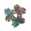
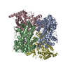
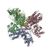
Journal: Mol Cell / Year: 2021
Title: A cooperative PNPase-Hfq-RNA carrier complex facilitates bacterial riboregulation.
Authors: Tom Dendooven / Dhriti Sinha / Alzbeta Roeselová / Todd A Cameron / Nicholas R De Lay / Ben F Luisi / Katarzyna J Bandyra /
Abstract: Polynucleotide phosphorylase (PNPase) is an ancient exoribonuclease conserved in the course of evolution and is found in species as diverse as bacteria and humans. Paradoxically, Escherichia coli ...Polynucleotide phosphorylase (PNPase) is an ancient exoribonuclease conserved in the course of evolution and is found in species as diverse as bacteria and humans. Paradoxically, Escherichia coli PNPase can act not only as an RNA degrading enzyme but also by an unknown mechanism as a chaperone for small regulatory RNAs (sRNAs), with pleiotropic consequences for gene regulation. We present structures of the ternary assembly formed by PNPase, the RNA chaperone Hfq, and sRNA and show that this complex boosts sRNA stability in vitro. Comparison of structures for PNPase in RNA carrier and degradation modes reveals how the RNA is rerouted away from the active site through interactions with Hfq and the KH and S1 domains. Together, these data explain how PNPase is repurposed to protect sRNAs from cellular ribonucleases such as RNase E and could aid RNA presentation to facilitate regulatory actions on target genes.
History
DepositionMay 6, 2021-
Header (metadata) releaseJul 7, 2021-
Map releaseJul 7, 2021-
UpdateJul 28, 2021-
Current statusJul 28, 2021Processing site: PDBe / Status: Released

-
Structure visualization

Movie
  • Surface view with section colored by density value
  • Surface level: 0.025
  • Imaged by UCSF Chimera
  • Download
  • Surface view colored by cylindrical radius
  • Surface level: 0.025
  • Imaged by UCSF Chimera
  • Download
  • Surface view with fitted model
  • Atomic models: PDB-7ogl
  • Surface level: 0.025
  • Imaged by UCSF Chimera
  • Download
Movie viewer
Structure viewerEM map:
SurfViewMolmilJmol/JSmol
Supplemental images

Downloads & links

-
Map

FileDownload / File: emd_12883.map.gz / Format: CCP4 / Size: 67 MB / Type: IMAGE STORED AS FLOATING POINT NUMBER (4 BYTES)
AnnotationSharpened map, b-factor -99
Projections & slices

Image control

Size
Brightness
Contrast
Others
AxesZ (Sec.)Y (Row.)X (Col.)
1.07 Å/pix.
x 260 pix.
= 276.9 Å
1.07 Å/pix.
x 260 pix.
= 276.9 Å
1.07 Å/pix.
x 260 pix.
= 276.9 Å

Surface

Projections

Slices (1/3)

Slices (1/2)

Slices (2/3)

Images are generated by Spider.

Voxel sizeX=Y=Z: 1.065 Å
Density
Contour LevelBy AUTHOR: 0.0337 / Movie #1: 0.025
Minimum - Maximum-0.11524918 - 0.16851738
Average (Standard dev.)-4.505516e-06 (±0.0050224597)
SymmetrySpace group: 1
Details

EMDB XML:

Map geometry
Axis orderXYZ
Origin000
Dimensions260260260
Spacing260260260
CellA=B=C: 276.90002 Å
α=β=γ: 90.0 °

CCP4 map header:

modeImage stored as Reals
Å/pix. X/Y/Z1.0651.0651.065
M x/y/z260260260
origin x/y/z0.0000.0000.000
length x/y/z276.900276.900276.900
α/β/γ90.00090.00090.000
start NX/NY/NZ192139186
NX/NY/NZ211274246
MAP C/R/S123
start NC/NR/NS000
NC/NR/NS260260260
D min/max/mean-0.1150.169-0.000

-
Supplemental data

-
Mask #1

Fileemd_12883_msk_1.map
Projections & Slices
AxesZYX

Projections

Slices (1/2)
Density Histograms

-
Additional map: Locally filtered map, b-factor -30

Fileemd_12883_additional_1.map
AnnotationLocally filtered map, b-factor -30
Projections & Slices
AxesZYX

Projections

Slices (1/2)
Density Histograms

-
Half map: Half map 1

Fileemd_12883_half_map_1.map
AnnotationHalf map 1
Projections & Slices
AxesZYX

Projections

Slices (1/2)
Density Histograms

-
Half map: Half map 2

Fileemd_12883_half_map_2.map
AnnotationHalf map 2
Projections & Slices
AxesZYX

Projections

Slices (1/2)
Density Histograms

-
Sample components

-
Entire : PNPase

EntireName: PNPase
Components
  • Complex: PNPase
    • Protein or peptide: Polyribonucleotide nucleotidyltransferasePolynucleotide phosphorylase
  • Ligand: water

-
Supramolecule #1: PNPase

SupramoleculeName: PNPase / type: complex / ID: 1 / Parent: 0 / Macromolecule list: #1
Source (natural)Organism: Escherichia coli (E. coli)
Recombinant expressionOrganism: Escherichia coli (E. coli)
Molecular weightTheoretical: 231 KDa

-
Macromolecule #1: Polyribonucleotide nucleotidyltransferase

MacromoleculeName: Polyribonucleotide nucleotidyltransferase / type: protein_or_peptide / ID: 1 / Number of copies: 3 / Enantiomer: LEVO / EC number: polyribonucleotide nucleotidyltransferase
Source (natural)Organism: Escherichia coli (strain K12) (bacteria) / Strain: K12
Molecular weightTheoretical: 77.189844 KDa
Recombinant expressionOrganism: Escherichia coli (E. coli)
SequenceString: MLNPIVRKFQ YGQHTVTLET GMMARQATAA VMVSMDDTAV FVTVVGQKKA KPGQDFFPLT VNYQERTYAA GRIPGSFFRR EGRPSEGET LIARLIDRPI RPLFPEGFVN EVQVIATVVS VNPQVNPDIV AMIGASAALS LSGIPFNGPI GAARVGYIND Q YVLNPTQD ...String:
MLNPIVRKFQ YGQHTVTLET GMMARQATAA VMVSMDDTAV FVTVVGQKKA KPGQDFFPLT VNYQERTYAA GRIPGSFFRR EGRPSEGET LIARLIDRPI RPLFPEGFVN EVQVIATVVS VNPQVNPDIV AMIGASAALS LSGIPFNGPI GAARVGYIND Q YVLNPTQD ELKESKLDLV VAGTEAAVLM VESEAQLLSE DQMLGAVVFG HEQQQVVIQN INELVKEAGK PRWDWQPEPV NE ALNARVA ALAEARLSDA YRITDKQERY AQVDVIKSET IATLLAEDET LDENELGEIL HAIEKNVVRS RVLAGEPRID GRE KDMIRG LDVRTGVLPR THGSALFTRG ETQALVTATL GTARDAQVLD ELMGERTDTF LFHYNFPPYS VGETGMVGSP KRRE IGHGR LAKRGVLAVM PDMDKFPYTV RVVSEITESN GSSSMASVCG ASLALMDAGV PIKAAVAGIA MGLVKEGDNY VVLSD ILGD EDHLGDMDFK VAGSRDGISA LQMDIKIEGI TKEIMQVALN QAKGARLHIL GVMEQAINAP RGDISEFAPR IHTIKI NPD KIKDVIGKGG SVIRALTEET GTTIEIEDDG TVKIAATDGE KAKHAIRRIE EITAEIEVGR VYTGKVTRIV DFGAFVA IG GGKEGLVHIS QIADKRVEKV TDYLQMGQEV PVKVLEVDRQ GRIRLSIKEA TEQSQPAAAP EAPAAEQGE

-
Macromolecule #2: water

MacromoleculeName: water / type: ligand / ID: 2 / Number of copies: 3 / Formula: HOH
Molecular weightTheoretical: 18.015 Da
Chemical component information

ChemComp-HOH:
WATER / Water

-
Experimental details

-
Structure determination

Methodcryo EM
Processingsingle particle reconstruction
Aggregation stateparticle

-
Sample preparation

BufferpH: 7.5
GridModel: UltrAuFoil R2/2 / Material: GOLD / Mesh: 300 / Pretreatment - Type: GLOW DISCHARGE
VitrificationCryogen name: ETHANE / Chamber humidity: 100 % / Chamber temperature: 277 K / Instrument: FEI VITROBOT MARK IV

-
Electron microscopy

MicroscopeFEI TITAN KRIOS
Electron beamAcceleration voltage: 300 kV / Electron source: FIELD EMISSION GUN
Electron opticsIllumination mode: FLOOD BEAM / Imaging mode: BRIGHT FIELDBright-field microscopy / Nominal defocus max: -0.0026000000000000003 µm / Nominal defocus min: -0.001 µm / Nominal magnification: 130000
Sample stageCooling holder cryogen: NITROGEN
Image recordingFilm or detector model: GATAN K2 SUMMIT (4k x 4k) / Detector mode: COUNTING / Digitization - Frames/image: 1-38 / Number grids imaged: 1 / Number real images: 1008 / Average exposure time: 12.0 sec. / Average electron dose: 53.0 e/Å2
Experimental equipment
Model: Titan Krios / Image courtesy: FEI Company

-
Image processing

CTF correctionSoftware: (Name: Gctf (ver. 1.06), Warp (ver. 1.0.9))
Startup modelType of model: NONE
Details: Initial maps were generated ab initio in Relion 3.1 via stochastic gradient descent
Initial angle assignmentType: RANDOM ASSIGNMENT / Software - Name: RELION (ver. 3.1)
Details: Initial maps were generated ab initio in Relion 3.1 via stochastic gradient descent
Final 3D classificationNumber classes: 6 / Software - Name: RELION (ver. 3.1)
Final angle assignmentType: MAXIMUM LIKELIHOOD / Software - Name: RELION (ver. 3.1)
Final reconstructionAlgorithm: BACK PROJECTION / Resolution.type: BY AUTHOR / Resolution: 3.4 Å / Resolution method: FSC 0.143 CUT-OFF / Software - Name: RELION (ver. 3.1) / Number images used: 58000
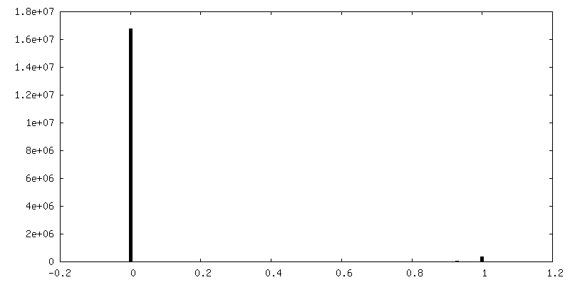
FSC plot (resolution estimation)

-
Atomic model buiding 1

RefinementProtocol: RIGID BODY FIT
Output model

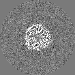
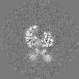
PDB-7ogl:
A cooperative PNPase-Hfq-RNA carrier complex facilitates bacterial riboregulation. apo-PNPase

+
About Yorodumi

-
News

-
Feb 9, 2022. New format data for meta-information of EMDB entries

New format data for meta-information of EMDB entries

  • Version 3 of the EMDB header file is now the official format.
  • The previous official version 1.9 will be removed from the archive.

Related info.:EMDB header

External links:wwPDB to switch to version 3 of the EMDB data model

-
Aug 12, 2020. Covid-19 info

Covid-19 info

URL: https://pdbj.org/emnavi/covid19.php

New page: Covid-19 featured information page in EM Navigator.

Related info.:Covid-19 info / Mar 5, 2020. Novel coronavirus structure data

+
Mar 5, 2020. Novel coronavirus structure data

Novel coronavirus structure data

Related info.:Yorodumi Speices / Aug 12, 2020. Covid-19 info

External links:COVID-19 featured content - PDBj / Molecule of the Month (242):Coronavirus Proteases

+
Jan 31, 2019. EMDB accession codes are about to change! (news from PDBe EMDB page)

EMDB accession codes are about to change! (news from PDBe EMDB page)

  • The allocation of 4 digits for EMDB accession codes will soon come to an end. Whilst these codes will remain in use, new EMDB accession codes will include an additional digit and will expand incrementally as the available range of codes is exhausted. The current 4-digit format prefixed with “EMD-” (i.e. EMD-XXXX) will advance to a 5-digit format (i.e. EMD-XXXXX), and so on. It is currently estimated that the 4-digit codes will be depleted around Spring 2019, at which point the 5-digit format will come into force.
  • The EM Navigator/Yorodumi systems omit the EMD- prefix.

Related info.:Q: What is EMD? / ID/Accession-code notation in Yorodumi/EM Navigator

External links:EMDB Accession Codes are Changing Soon! / Contact to PDBj

+
Jul 12, 2017. Major update of PDB

Major update of PDB

  • wwPDB released updated PDB data conforming to the new PDBx/mmCIF dictionary.
  • This is a major update changing the version number from 4 to 5, and with Remediation, in which all the entries are updated.
  • In this update, many items about electron microscopy experimental information are reorganized (e.g. em_software).
  • Now, EM Navigator and Yorodumi are based on the updated data.

External links:wwPDB Remediation / Enriched Model Files Conforming to OneDep Data Standards Now Available in the PDB FTP Archive

-
Yorodumi

Thousand views of thousand structures

  • Yorodumi is a browser for structure data from EMDB, PDB, SASBDB, etc.
  • This page is also the successor to EM Navigator detail page, and also detail information page/front-end page for Omokage search.
  • The word "yorodu" (or yorozu) is an old Japanese word meaning "ten thousand". "mi" (miru) is to see.

Related info.:EMDB / PDB / SASBDB / Comparison of 3 databanks / Yorodumi Search / Aug 31, 2016. New EM Navigator & Yorodumi / Yorodumi Papers / Jmol/JSmol / Function and homology information / Changes in new EM Navigator and Yorodumi

Read more