[English] 日本語
Yorodumi
- EMDB-12876: Cryo-EM map of Scc2 bound to cohesin trimer -

+
Open data


ID or keywords:

Loading...

-
Basic information

Entry
Database: EMDB / ID: EMD-12876
TitleCryo-EM map of Scc2 bound to cohesin trimer
Map dataScc2 bound to cohesin trimer
Sample
  • Complex: Cohesin
Function / homology
Function and homology information


Establishment of Sister Chromatid Cohesion / Resolution of Sister Chromatid Cohesion / cohesin loader activity / meiotic cohesin complex / establishment of meiotic sister chromatid cohesion / mitotic cohesin complex / DNA secondary structure binding / cohesin complex / synaptonemal complex assembly / SUMOylation of DNA damage response and repair proteins ...Establishment of Sister Chromatid Cohesion / Resolution of Sister Chromatid Cohesion / cohesin loader activity / meiotic cohesin complex / establishment of meiotic sister chromatid cohesion / mitotic cohesin complex / DNA secondary structure binding / cohesin complex / synaptonemal complex assembly / SUMOylation of DNA damage response and repair proteins / meiotic sister chromatid cohesion / replication-born double-strand break repair via sister chromatid exchange / establishment of mitotic sister chromatid cohesion / reciprocal meiotic recombination / sister chromatid cohesion / mitotic sister chromatid cohesion / minor groove of adenine-thymine-rich DNA binding / mitotic sister chromatid segregation / double-strand break repair / double-stranded DNA binding / cell division / protein kinase binding / ATP hydrolysis activity / DNA binding / ATP binding / nucleus / identical protein binding
Similarity search - Function
Smc1, ATP-binding cassette domain / Structural maintenance of chromosomes 3, ABC domain, eukaryotic / Structural maintenance of chromosomes protein / SMCs flexible hinge / SMCs flexible hinge superfamily / SMC proteins Flexible Hinge Domain / SMC proteins Flexible Hinge Domain / RecF/RecN/SMC, N-terminal / RecF/RecN/SMC N terminal domain / P-loop containing nucleoside triphosphate hydrolase
Similarity search - Domain/homology
Structural maintenance of chromosomes protein 1 / Structural maintenance of chromosomes protein 3
Similarity search - Component
Biological speciesSaccharomyces cerevisiae (brewer's yeast)
Methodsingle particle reconstruction / cryo EM / Resolution: 13.0 Å
AuthorsGonzalez Llamazares A / Lee B / Lowe J
Citation
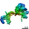
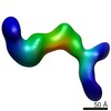
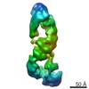
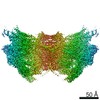
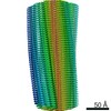
Journal: Elife / Year: 2021
Title: Folding of cohesin's coiled coil is important for Scc2/4-induced association with chromosomes.
Authors: Naomi J Petela / Andres Gonzalez Llamazares / Sarah Dixon / Bin Hu / Byung-Gil Lee / Jean Metson / Heekyo Seo / Antonio Ferrer-Harding / Menelaos Voulgaris / Thomas Gligoris / James Collier ...Authors: Naomi J Petela / Andres Gonzalez Llamazares / Sarah Dixon / Bin Hu / Byung-Gil Lee / Jean Metson / Heekyo Seo / Antonio Ferrer-Harding / Menelaos Voulgaris / Thomas Gligoris / James Collier / Byung-Ha Oh / Jan Löwe / Kim A Nasmyth /
Abstract: Cohesin's association with and translocation along chromosomal DNAs depend on an ATP hydrolysis cycle driving the association and subsequent release of DNA. This involves DNA being 'clamped' by Scc2 ...Cohesin's association with and translocation along chromosomal DNAs depend on an ATP hydrolysis cycle driving the association and subsequent release of DNA. This involves DNA being 'clamped' by Scc2 and ATP-dependent engagement of cohesin's Smc1 and Smc3 head domains. Scc2's replacement by Pds5 abrogates cohesin's ATPase and has an important role in halting DNA loop extrusion. The ATPase domains of all SMC proteins are separated from their hinge dimerisation domains by 50-nm-long coiled coils, which have been observed to zip up along their entire length and fold around an elbow, thereby greatly shortening the distance between hinges and ATPase heads. Whether folding exists in vivo or has any physiological importance is not known. We present here a cryo-EM structure of the form of cohesin that reveals the structure of folded and zipped-up coils in unprecedented detail and shows that Scc2 can associate with Smc1's ATPase head even when it is fully disengaged from that of Smc3. Using cysteine-specific crosslinking, we show that cohesin's coiled coils are frequently folded in vivo, including when cohesin holds sister chromatids together. Moreover, we describe a mutation () within Smc1's hinge that alters how Scc2 and Pds5 interact with Smc1's hinge and that enables Scc2 to support loading in the absence of its normal partner Scc4. The mutant phenotype of loading without Scc4 is only explicable if loading depends on an association between Scc2/4 and cohesin's hinge, which in turn requires coiled coil folding.
History
DepositionMay 5, 2021-
Header (metadata) releaseJul 28, 2021-
Map releaseJul 28, 2021-
UpdateJul 28, 2021-
Current statusJul 28, 2021Processing site: PDBe / Status: Released

-
Structure visualization
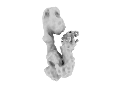
Movie
  • Surface view with section colored by density value
  • Surface level: 0.06
  • Imaged by UCSF Chimera
  • Download
  • Surface view colored by radius
  • Surface level: 0.06
  • Imaged by UCSF Chimera
  • Download
Movie viewer
Structure viewerEM map:
SurfViewMolmilJmol/JSmol
Supplemental images

Downloads & links

-
Map

FileDownload / File: emd_12876.map.gz / Format: CCP4 / Size: 30.5 MB / Type: IMAGE STORED AS FLOATING POINT NUMBER (4 BYTES)
AnnotationScc2 bound to cohesin trimer
Voxel sizeX=Y=Z: 1.72 Å
Density
Contour LevelBy AUTHOR: 0.06 / Movie #1: 0.06
Minimum - Maximum-0.26036233 - 0.7627526
Average (Standard dev.)0.0011641289 (±0.041564826)
SymmetrySpace group: 1
Details

EMDB XML:

Map geometry
Axis orderXYZ
Origin000
Dimensions200200200
Spacing200200200
CellA=B=C: 344.0 Å
α=β=γ: 90.0 °

CCP4 map header:

modeImage stored as Reals
Å/pix. X/Y/Z1.721.721.72
M x/y/z200200200
origin x/y/z0.0000.0000.000
length x/y/z344.000344.000344.000
α/β/γ90.00090.00090.000
start NX/NY/NZ1331310
NX/NY/NZ223226424
MAP C/R/S123
start NC/NR/NS000
NC/NR/NS200200200
D min/max/mean-0.2600.7630.001

-
Supplemental data

-
Sample components

-
Entire : Cohesin

EntireName: Cohesin
Components
  • Complex: Cohesin

-
Supramolecule #1: Cohesin

SupramoleculeName: Cohesin / type: complex / ID: 1 / Parent: 0
Source (natural)Organism: Saccharomyces cerevisiae (brewer's yeast)
Recombinant expressionOrganism: Insect cell expression vector pTIE1 (others)

-
Experimental details

-
Structure determination

Methodcryo EM
Processingsingle particle reconstruction
Aggregation stateparticle

-
Sample preparation

BufferpH: 7.5
VitrificationCryogen name: ETHANE

-
Electron microscopy

MicroscopeTFS KRIOS
Electron beamAcceleration voltage: 300 kV / Electron source: FIELD EMISSION GUN
Electron opticsIllumination mode: FLOOD BEAM / Imaging mode: BRIGHT FIELDBright-field microscopy
Image recordingFilm or detector model: GATAN K2 SUMMIT (4k x 4k) / Average electron dose: 42.5 e/Å2
Experimental equipment
Model: Titan Krios / Image courtesy: FEI Company

-
Image processing

Initial angle assignmentType: MAXIMUM LIKELIHOOD
Final angle assignmentType: MAXIMUM LIKELIHOOD
Final reconstructionResolution.type: BY AUTHOR / Resolution: 13.0 Å / Resolution method: FSC 0.143 CUT-OFF / Number images used: 32491

+
About Yorodumi

-
News

-
Feb 9, 2022. New format data for meta-information of EMDB entries

New format data for meta-information of EMDB entries

  • Version 3 of the EMDB header file is now the official format.
  • The previous official version 1.9 will be removed from the archive.

Related info.:EMDB header

External links:wwPDB to switch to version 3 of the EMDB data model

-
Aug 12, 2020. Covid-19 info

Covid-19 info

URL: https://pdbj.org/emnavi/covid19.php

New page: Covid-19 featured information page in EM Navigator.

Related info.:Covid-19 info / Mar 5, 2020. Novel coronavirus structure data

+
Mar 5, 2020. Novel coronavirus structure data

Novel coronavirus structure data

Related info.:Yorodumi Speices / Aug 12, 2020. Covid-19 info

External links:COVID-19 featured content - PDBj / Molecule of the Month (242):Coronavirus Proteases

+
Jan 31, 2019. EMDB accession codes are about to change! (news from PDBe EMDB page)

EMDB accession codes are about to change! (news from PDBe EMDB page)

  • The allocation of 4 digits for EMDB accession codes will soon come to an end. Whilst these codes will remain in use, new EMDB accession codes will include an additional digit and will expand incrementally as the available range of codes is exhausted. The current 4-digit format prefixed with “EMD-” (i.e. EMD-XXXX) will advance to a 5-digit format (i.e. EMD-XXXXX), and so on. It is currently estimated that the 4-digit codes will be depleted around Spring 2019, at which point the 5-digit format will come into force.
  • The EM Navigator/Yorodumi systems omit the EMD- prefix.

Related info.:Q: What is EMD? / ID/Accession-code notation in Yorodumi/EM Navigator

External links:EMDB Accession Codes are Changing Soon! / Contact to PDBj

+
Jul 12, 2017. Major update of PDB

Major update of PDB

  • wwPDB released updated PDB data conforming to the new PDBx/mmCIF dictionary.
  • This is a major update changing the version number from 4 to 5, and with Remediation, in which all the entries are updated.
  • In this update, many items about electron microscopy experimental information are reorganized (e.g. em_software).
  • Now, EM Navigator and Yorodumi are based on the updated data.

External links:wwPDB Remediation / Enriched Model Files Conforming to OneDep Data Standards Now Available in the PDB FTP Archive

-
Yorodumi

Thousand views of thousand structures

  • Yorodumi is a browser for structure data from EMDB, PDB, SASBDB, etc.
  • This page is also the successor to EM Navigator detail page, and also detail information page/front-end page for Omokage search.
  • The word "yorodu" (or yorozu) is an old Japanese word meaning "ten thousand". "mi" (miru) is to see.

Related info.:EMDB / PDB / SASBDB / Comparison of 3 databanks / Yorodumi Search / Aug 31, 2016. New EM Navigator & Yorodumi / Yorodumi Papers / Jmol/JSmol / Function and homology information / Changes in new EM Navigator and Yorodumi

Read more