+ Open data
- Basic information
| Entry | Database: EMDB / ID: EMD-6437 | |||||||||
|---|---|---|---|---|---|---|---|---|---|---|
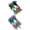
| Title | RCT reconstruction of CMV protein complex | |||||||||
 Map data Map data | RCT reconstruction of CMV protein complex | |||||||||
 Sample Sample |
| |||||||||
| Biological species |  Cytomegalovirus / unidentified (others) Cytomegalovirus / unidentified (others) | |||||||||
| Method | single particle reconstruction / Resolution: 30.0 Å | |||||||||
 Authors Authors | Ciferri C / Cianfrocco MA / Chandramouli S / Carfi A | |||||||||
 Citation Citation |  Journal: PLoS Pathog / Year: 2015 Journal: PLoS Pathog / Year: 2015Title: Antigenic Characterization of the HCMV gH/gL/gO and Pentamer Cell Entry Complexes Reveals Binding Sites for Potently Neutralizing Human Antibodies. Authors: Claudio Ciferri / Sumana Chandramouli / Alexander Leitner / Danilo Donnarumma / Michael A Cianfrocco / Rachel Gerrein / Kristian Friedrich / Yukti Aggarwal / Giuseppe Palladino / Ruedi ...Authors: Claudio Ciferri / Sumana Chandramouli / Alexander Leitner / Danilo Donnarumma / Michael A Cianfrocco / Rachel Gerrein / Kristian Friedrich / Yukti Aggarwal / Giuseppe Palladino / Ruedi Aebersold / Nathalie Norais / Ethan C Settembre / Andrea Carfi /    Abstract: Human Cytomegalovirus (HCMV) is a major cause of morbidity and mortality in transplant patients and in fetuses following congenital infection. The glycoprotein complexes gH/gL/gO and ...Human Cytomegalovirus (HCMV) is a major cause of morbidity and mortality in transplant patients and in fetuses following congenital infection. The glycoprotein complexes gH/gL/gO and gH/gL/UL128/UL130/UL131A (Pentamer) are required for HCMV entry in fibroblasts and endothelial/epithelial cells, respectively, and are targeted by potently neutralizing antibodies in the infected host. Using purified soluble forms of gH/gL/gO and Pentamer as well as a panel of naturally elicited human monoclonal antibodies, we determined the location of key neutralizing epitopes on the gH/gL/gO and Pentamer surfaces. Mass Spectrometry (MS) coupled to Chemical Crosslinking or to Hydrogen Deuterium Exchange was used to define residues that are either in proximity or part of neutralizing epitopes on the glycoprotein complexes. We also determined the molecular architecture of the gH/gL/gO- and Pentamer-antibody complexes by Electron Microscopy (EM) and 3D reconstructions. The EM analysis revealed that the Pentamer specific neutralizing antibodies bind to two opposite surfaces of the complex, suggesting that they may neutralize infection by different mechanisms. Together, our data identify the location of neutralizing antibodies binding sites on the gH/gL/gO and Pentamer complexes and provide a framework for the development of antibodies and vaccines against HCMV. | |||||||||
| History |
|
- Structure visualization
| Movie |
 Movie viewer Movie viewer |
|---|---|
| Structure viewer | EM map:  SurfView SurfView Molmil Molmil Jmol/JSmol Jmol/JSmol |
| Supplemental images |
- Downloads & links
-EMDB archive
| Map data |  emd_6437.map.gz emd_6437.map.gz | 3.4 MB |  EMDB map data format EMDB map data format | |
|---|---|---|---|---|
| Header (meta data) |  emd-6437-v30.xml emd-6437-v30.xml emd-6437.xml emd-6437.xml | 7.9 KB 7.9 KB | Display Display |  EMDB header EMDB header |
| Images |  emd_6437.jpg emd_6437.jpg | 15.6 KB | ||
| Archive directory |  http://ftp.pdbj.org/pub/emdb/structures/EMD-6437 http://ftp.pdbj.org/pub/emdb/structures/EMD-6437 ftp://ftp.pdbj.org/pub/emdb/structures/EMD-6437 ftp://ftp.pdbj.org/pub/emdb/structures/EMD-6437 | HTTPS FTP |
-Validation report
| Summary document |  emd_6437_validation.pdf.gz emd_6437_validation.pdf.gz | 77.2 KB | Display |  EMDB validaton report EMDB validaton report |
|---|---|---|---|---|
| Full document |  emd_6437_full_validation.pdf.gz emd_6437_full_validation.pdf.gz | 76.3 KB | Display | |
| Data in XML |  emd_6437_validation.xml.gz emd_6437_validation.xml.gz | 495 B | Display | |
| Arichive directory |  https://ftp.pdbj.org/pub/emdb/validation_reports/EMD-6437 https://ftp.pdbj.org/pub/emdb/validation_reports/EMD-6437 ftp://ftp.pdbj.org/pub/emdb/validation_reports/EMD-6437 ftp://ftp.pdbj.org/pub/emdb/validation_reports/EMD-6437 | HTTPS FTP |
-Related structure data
- Links
Links
| EMDB pages |  EMDB (EBI/PDBe) / EMDB (EBI/PDBe) /  EMDataResource EMDataResource |
|---|
- Map
Map
| File |  Download / File: emd_6437.map.gz / Format: CCP4 / Size: 3.7 MB / Type: IMAGE STORED AS FLOATING POINT NUMBER (4 BYTES) Download / File: emd_6437.map.gz / Format: CCP4 / Size: 3.7 MB / Type: IMAGE STORED AS FLOATING POINT NUMBER (4 BYTES) | ||||||||||||||||||||||||||||||||||||||||||||||||||||||||||||||||||||
|---|---|---|---|---|---|---|---|---|---|---|---|---|---|---|---|---|---|---|---|---|---|---|---|---|---|---|---|---|---|---|---|---|---|---|---|---|---|---|---|---|---|---|---|---|---|---|---|---|---|---|---|---|---|---|---|---|---|---|---|---|---|---|---|---|---|---|---|---|---|
| Annotation | RCT reconstruction of CMV protein complex | ||||||||||||||||||||||||||||||||||||||||||||||||||||||||||||||||||||
| Projections & slices | Image control
Images are generated by Spider. | ||||||||||||||||||||||||||||||||||||||||||||||||||||||||||||||||||||
| Voxel size | X=Y=Z: 4.3 Å | ||||||||||||||||||||||||||||||||||||||||||||||||||||||||||||||||||||
| Density |
| ||||||||||||||||||||||||||||||||||||||||||||||||||||||||||||||||||||
| Symmetry | Space group: 1 | ||||||||||||||||||||||||||||||||||||||||||||||||||||||||||||||||||||
| Details | EMDB XML:
CCP4 map header:
| ||||||||||||||||||||||||||||||||||||||||||||||||||||||||||||||||||||
-Supplemental data
- Sample components
-Entire : RCT reconstruction of CMV Complex bound to neutralizing Fabs
| Entire | Name: RCT reconstruction of CMV Complex bound to neutralizing Fabs |
|---|---|
| Components |
|
-Supramolecule #1000: RCT reconstruction of CMV Complex bound to neutralizing Fabs
| Supramolecule | Name: RCT reconstruction of CMV Complex bound to neutralizing Fabs type: sample / ID: 1000 / Number unique components: 2 |
|---|
-Macromolecule #1: CMV protein complex
| Macromolecule | Name: CMV protein complex / type: protein_or_peptide / ID: 1 / Recombinant expression: Yes |
|---|---|
| Source (natural) | Organism:  Cytomegalovirus Cytomegalovirus |
| Recombinant expression | Organism:  Homo sapiens (human) Homo sapiens (human) |
-Macromolecule #2: neutralizing antibody
| Macromolecule | Name: neutralizing antibody / type: protein_or_peptide / ID: 2 / Recombinant expression: No / Database: NCBI |
|---|---|
| Source (natural) | Organism: unidentified (others) |
-Experimental details
-Structure determination
 Processing Processing | single particle reconstruction |
|---|---|
| Aggregation state | particle |
- Sample preparation
| Vitrification | Cryogen name: NONE / Instrument: OTHER |
|---|
- Electron microscopy
| Microscope | FEI TECNAI 12 |
|---|---|
| Date | Jun 1, 2014 |
| Image recording | Category: CCD / Film or detector model: FEI CETA (4k x 4k) |
| Electron beam | Acceleration voltage: 120 kV / Electron source: LAB6 |
| Electron optics | Illumination mode: FLOOD BEAM / Imaging mode: BRIGHT FIELD |
| Sample stage | Specimen holder model: SIDE ENTRY, EUCENTRIC |
- Image processing
| Final reconstruction | Algorithm: OTHER / Resolution.type: BY AUTHOR / Resolution: 30.0 Å / Resolution method: OTHER / Number images used: 2000 |
|---|
 Movie
Movie Controller


 UCSF Chimera


 Z (Sec.)
 Y (Row.)
 X (Col.)


