 ムービー
ムービー コントローラー


+ データを開く

- 基本情報
基本情報
| 登録情報 | データベース: EMDB / ID: EMD-1281 | |||||||||
|---|---|---|---|---|---|---|---|---|---|---|
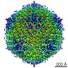
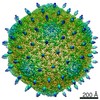
| タイトル | Determinants of bacteriophage phi29 head morphology. | |||||||||
 マップデータ マップデータ | This map is a cryo-EM 3D reconstruction of a fibered, T=4 isometric variant of bacteriophage phi29 | |||||||||
 試料 試料 |
| |||||||||
| 生物種 |   Bacillus phage phi29 (ファージ) Bacillus phage phi29 (ファージ) | |||||||||
| 手法 | 単粒子再構成法 / クライオ電子顕微鏡法 / 解像度: 20.0 Å | |||||||||
 データ登録者 データ登録者 | Choi KH / Morais MC / Anderson DL / Rossmann MG | |||||||||
 引用 引用 | ジャーナル: Structure / 年: 2006 タイトル: Determinants of bacteriophage phi29 head morphology. 著者: Kyung H Choi / Marc C Morais / Dwight L Anderson / Michael G Rossmann /  要旨: Bacteriophage phi29 requires scaffolding protein to assemble the 450 x 540 A prolate prohead with T = 3 symmetry end caps. In infections with a temperature-sensitive mutant scaffolding protein, ...Bacteriophage phi29 requires scaffolding protein to assemble the 450 x 540 A prolate prohead with T = 3 symmetry end caps. In infections with a temperature-sensitive mutant scaffolding protein, capsids assemble predominantly into 370 A diameter isometric particles with T = 3 symmetry that lack a head-tail connector. However, a few larger, 430 A diameter, particles are also assembled. Cryo-electron microscopy shows that these larger particles are icosahedral with T = 4 symmetry. The prolate prohead, as well as the two isometric capsids with T = 3 and T = 4 symmetry, are composed of similar pentamers and differently skewed hexamers. The skewing of the hexamers in the equatorial region of proheads and in the T = 4 isometric particles reflects their different environments. One of the functions of the scaffolding protein, present in the prohead, may be to stabilize skewed hexamers during assembly. | |||||||||
| 履歴 |
|
- 構造の表示
構造の表示
| ムービー |
 ムービービューア ムービービューア |
|---|---|
| 構造ビューア | EMマップ: SurfViewMolmilJmol/JSmol |
| 添付画像 |

- ダウンロードとリンク
-EMDBアーカイブ
| マップデータ | emd_1281.map.gz | 106.1 MB | EMDBマップデータ形式 | |
|---|---|---|---|---|
| ヘッダ (付随情報) | emd-1281-v30.xmlemd-1281.xml | 8.4 KB 8.4 KB | 表示 表示 | EMDBヘッダ |
| 画像 | 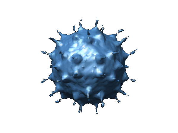 1281.gif 1281.gif | 46.3 KB | ||
| アーカイブディレクトリ |  http://ftp.pdbj.org/pub/emdb/structures/EMD-1281ftp://ftp.pdbj.org/pub/emdb/structures/EMD-1281 http://ftp.pdbj.org/pub/emdb/structures/EMD-1281ftp://ftp.pdbj.org/pub/emdb/structures/EMD-1281 | HTTPS FTP |
-検証レポート
| 文書・要旨 | emd_1281_validation.pdf.gz | 215.7 KB | 表示 | EMDB検証レポート |
|---|---|---|---|---|
| 文書・詳細版 | emd_1281_full_validation.pdf.gz | 214.9 KB | 表示 | |
| XML形式データ | emd_1281_validation.xml.gz | 7.9 KB | 表示 | |
| アーカイブディレクトリ | https://ftp.pdbj.org/pub/emdb/validation_reports/EMD-1281ftp://ftp.pdbj.org/pub/emdb/validation_reports/EMD-1281 | HTTPS FTP |
-関連構造データ


-リンク
| EMDBのページ | EMDB (EBI/PDBe) / EMDataResource |
|---|
-マップ
| ファイル | ダウンロード / ファイル: emd_1281.map.gz / 形式: CCP4 / 大きさ: 276 MB / タイプ: IMAGE STORED AS FLOATING POINT NUMBER (4 BYTES) | ||||||||||||||||||||||||||||||||||||||||||||||||||||||||||||||||||||
|---|---|---|---|---|---|---|---|---|---|---|---|---|---|---|---|---|---|---|---|---|---|---|---|---|---|---|---|---|---|---|---|---|---|---|---|---|---|---|---|---|---|---|---|---|---|---|---|---|---|---|---|---|---|---|---|---|---|---|---|---|---|---|---|---|---|---|---|---|---|
| 注釈 | This map is a cryo-EM 3D reconstruction of a fibered, T=4 isometric variant of bacteriophage phi29 | ||||||||||||||||||||||||||||||||||||||||||||||||||||||||||||||||||||
| 投影像・断面図 | 画像のコントロール
画像は Spider により作成 | ||||||||||||||||||||||||||||||||||||||||||||||||||||||||||||||||||||
| ボクセルのサイズ | X=Y=Z: 3.68 Å | ||||||||||||||||||||||||||||||||||||||||||||||||||||||||||||||||||||
| 密度 |
| ||||||||||||||||||||||||||||||||||||||||||||||||||||||||||||||||||||
| 対称性 | 空間群: 1 | ||||||||||||||||||||||||||||||||||||||||||||||||||||||||||||||||||||
| 詳細 | EMDB XML:
CCP4マップ ヘッダ情報:
| ||||||||||||||||||||||||||||||||||||||||||||||||||||||||||||||||||||
 Z (Sec.)
 Y (Row.)
 X (Col.)


-添付データ
- 試料の構成要素
-全体 : fibered isometric variants of bacteriophage phi29
| 全体 | 名称: fibered isometric variants of bacteriophage phi29 |
|---|---|
| 要素 |
|
-超分子 #1000: fibered isometric variants of bacteriophage phi29
| 超分子 | 名称: fibered isometric variants of bacteriophage phi29 / タイプ: sample / ID: 1000 / 集合状態: T4 icosahderal / Number unique components: 1 |
|---|
-超分子 #1: Bacillus phage phi29
| 超分子 | 名称: Bacillus phage phi29 / タイプ: virus / ID: 1 / Name.synonym: phi29 / NCBI-ID: 10756 / 生物種: Bacillus phage phi29 / ウイルスタイプ: VIRUS-LIKE PARTICLE / ウイルス・単離状態: SPECIES / ウイルス・エンベロープ: No / ウイルス・中空状態: Yes / Syn species name: phi29 |
|---|---|
| 宿主 | 生物種:  |
| 分子量 | 理論値: 17.3 MDa |
| ウイルス殻 | Shell ID: 1 / 名称: gp8 / 直径: 430 Å / T番号(三角分割数): 4 |
-実験情報
-構造解析
| 手法 | クライオ電子顕微鏡法 |
|---|---|
 解析 解析 | 単粒子再構成法 |
| 試料の集合状態 | particle |
-試料調製
| 緩衝液 | pH: 7.8 / 詳細: 25 mM Tris 5 mM MgCl2 50 mM NaCl 2 mM sodium azide |
|---|---|
| グリッド | 詳細: holey carbon |
| 凍結 | 凍結剤: ETHANE / チャンバー内温度: 113 K |
- 電子顕微鏡法
| 顕微鏡 | FEI/PHILIPS CM200FEG |
|---|---|
| 日付 | 1997年10月10日 |
| 撮影 | カテゴリ: FILM / フィルム・検出器のモデル: KODAK SO-163 FILM / デジタル化 - スキャナー: ZEISS SCAI / デジタル化 - サンプリング間隔: 3.68 µm / 実像数: 28 / 平均電子線量: 10 e/Å2 / ビット/ピクセル: 8 |
| Tilt angle min | 0 |
| Tilt angle max | 0 |
| 電子線 | 加速電圧: 200 kV / 電子線源:  FIELD EMISSION GUN FIELD EMISSION GUN |
| 電子光学系 | 照射モード: FLOOD BEAM / 撮影モード: BRIGHT FIELD / Cs: 2.0 mm / 最大 デフォーカス(公称値): 3.903 µm / 最小 デフォーカス(公称値): 1.091 µm / 倍率(公称値): 38000 |
| 試料ステージ | 試料ホルダー: Side entry liquid nitrogen-cooled cryo specimen holder 試料ホルダーモデル: GATAN LIQUID NITROGEN |
-画像解析
| CTF補正 | 詳細: CTF correction, phases and amplitudes, for each micrograph |
|---|---|
| 最終 再構成 | 想定した対称性 - 点群: I (正20面体型対称) / アルゴリズム: OTHER / 解像度のタイプ: BY AUTHOR / 解像度: 20.0 Å / 解像度の算出法: FSC 0.5 CUT-OFF / ソフトウェア - 名称: EMAN PFT P3DR POR / 使用した粒子像数: 785 |
