 Movie
Movie Controller

[English] 日本語
 Yorodumi
- EMDB-1280: Cryo-electron microscopy study of bacteriophage T4 displaying ant... -
+ Open data

- Basic information
| Entry | Database: EMDB / ID: EMD-1280 | |||||||||
|---|---|---|---|---|---|---|---|---|---|---|
| Title | Cryo-electron microscopy study of bacteriophage T4 displaying anthrax toxin proteins. | |||||||||
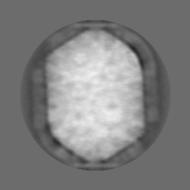
 Map data Map data | Bacteriophage T4 capsid decorated with anthrax toxin proteins. N-terminal domain of the anhrax lethal factor (nLF) was fused with T4 Soc protein. nLF-Soc molecules were attached to the surface of Hoc-minus Soc-minus T4 phage mutant. Then the anthrax protective antigen, PA63, heptamers were attached to the capsid-exposed LFn domains. | |||||||||
 Sample Sample |
| |||||||||
| Biological species |   Enterobacteria phage T4 (virus) Enterobacteria phage T4 (virus) | |||||||||
| Method | single particle reconstruction / cryo EM / Resolution: 35.0 Å | |||||||||
 Authors Authors | Fokine A / Bowman VD / Battisti AJ / Li Q / Chipman PR / Rao VB / Rossmann MG | |||||||||
 Citation Citation | Journal: Virology / Year: 2007 Title: Cryo-electron microscopy study of bacteriophage T4 displaying anthrax toxin proteins. Authors: Andrei Fokine / Valorie D Bowman / Anthony J Battisti / Qin Li / Paul R Chipman / Venigalla B Rao / Michael G Rossmann /  Abstract: The bacteriophage T4 capsid contains two accessory surface proteins, the small outer capsid protein (Soc, 870 copies) and the highly antigenic outer capsid protein (Hoc, 155 copies). As these are ...The bacteriophage T4 capsid contains two accessory surface proteins, the small outer capsid protein (Soc, 870 copies) and the highly antigenic outer capsid protein (Hoc, 155 copies). As these are dispensable for capsid formation, they can be used for displaying proteins and macromolecular complexes on the T4 capsid surface. Anthrax toxin components were attached to the T4 capsid as a fusion protein of the N-terminal domain of the anthrax lethal factor (LFn) with Soc. The LFn-Soc fusion protein was complexed in vitro with Hoc(-)Soc(-)T4 phage. Subsequently, cleaved anthrax protective antigen heptamers (PA63)(7) were attached to the exposed LFn domains. A cryo-electron microscopy study of the decorated T4 particles shows the complex of PA63 heptamers with LFn-Soc on the phage surface. Although the cryo-electron microscopy reconstruction is unable to differentiate on its own between different proposed models of the anthrax toxin, the density is consistent with a model that had predicted the orientation and position of three LFn molecules bound to one PA63 heptamer. | |||||||||
| History |
|
- Structure visualization
| Movie |
 Movie viewer Movie viewer |
|---|---|
| Structure viewer | EM map: SurfViewMolmilJmol/JSmol |
| Supplemental images |
- Downloads & links
-EMDB archive
| Map data | emd_1280.map.gz | 19.6 MB | EMDB map data format | |
|---|---|---|---|---|
| Header (meta data) | emd-1280-v30.xmlemd-1280.xml | 10.9 KB 10.9 KB | Display Display | EMDB header |
| Images |  1280.gif 1280.gif | 148.7 KB | ||
| Archive directory |  http://ftp.pdbj.org/pub/emdb/structures/EMD-1280ftp://ftp.pdbj.org/pub/emdb/structures/EMD-1280 http://ftp.pdbj.org/pub/emdb/structures/EMD-1280ftp://ftp.pdbj.org/pub/emdb/structures/EMD-1280 | HTTPS FTP |
-Related structure data
| Similar structure data |
|---|


-Links
| EMDB pages | EMDB (EBI/PDBe) / EMDataResource |
|---|
-Map
| File | Download / File: emd_1280.map.gz / Format: CCP4 / Size: 78.3 MB / Type: IMAGE STORED AS FLOATING POINT NUMBER (4 BYTES) | ||||||||||||||||||||||||||||||||||||||||||||||||||||||||||||||||||||
|---|---|---|---|---|---|---|---|---|---|---|---|---|---|---|---|---|---|---|---|---|---|---|---|---|---|---|---|---|---|---|---|---|---|---|---|---|---|---|---|---|---|---|---|---|---|---|---|---|---|---|---|---|---|---|---|---|---|---|---|---|---|---|---|---|---|---|---|---|---|
| Annotation | Bacteriophage T4 capsid decorated with anthrax toxin proteins. N-terminal domain of the anhrax lethal factor (nLF) was fused with T4 Soc protein. nLF-Soc molecules were attached to the surface of Hoc-minus Soc-minus T4 phage mutant. Then the anthrax protective antigen, PA63, heptamers were attached to the capsid-exposed LFn domains. | ||||||||||||||||||||||||||||||||||||||||||||||||||||||||||||||||||||
| Projections & slices | Image control
Images are generated by Spider. | ||||||||||||||||||||||||||||||||||||||||||||||||||||||||||||||||||||
| Voxel size | X=Y=Z: 5.96 Å | ||||||||||||||||||||||||||||||||||||||||||||||||||||||||||||||||||||
| Density |
| ||||||||||||||||||||||||||||||||||||||||||||||||||||||||||||||||||||
| Symmetry | Space group: 1 | ||||||||||||||||||||||||||||||||||||||||||||||||||||||||||||||||||||
| Details | EMDB XML:
CCP4 map header:
| ||||||||||||||||||||||||||||||||||||||||||||||||||||||||||||||||||||
 Z (Sec.)
 Y (Row.)
 X (Col.)


-Supplemental data
- Sample components
-Entire : Bacteriophage T4 decorated with anthrax toxin proteins
| Entire | Name: Bacteriophage T4 decorated with anthrax toxin proteins |
|---|---|
| Components |
|
-Supramolecule #1000: Bacteriophage T4 decorated with anthrax toxin proteins
| Supramolecule | Name: Bacteriophage T4 decorated with anthrax toxin proteins type: sample / ID: 1000 Details: Bacteriophage T4 decorated with anthrax toxin proteins. N-terminal domain of the anhrax lethal factor (nLF) was fused with T4 Soc protein. nLF-Soc molecules were attached to the surface of ...Details: Bacteriophage T4 decorated with anthrax toxin proteins. N-terminal domain of the anhrax lethal factor (nLF) was fused with T4 Soc protein. nLF-Soc molecules were attached to the surface of Hoc-minus Soc-minus T4 phage mutant. Then the anthrax protective antigen, PA63, heptamers were attached to the capsid-exposed LFn domains. Number unique components: 3 |
|---|
-Supramolecule #1: Enterobacteria phage T4
| Supramolecule | Name: Enterobacteria phage T4 / type: virus / ID: 1 / NCBI-ID: 10665 / Sci species name: Enterobacteria phage T4 / Virus type: VIRION / Virus isolate: STRAIN / Virus enveloped: No / Virus empty: No |
|---|---|
| Host (natural) | Organism: |
| Virus shell | Shell ID: 1 / Name: capsid shell / Diameter: 1200 Å / T number (triangulation number): 20 |
| Virus shell | Shell ID: 2 / Diameter: 850 Å / T number (triangulation number): 13 |
-Macromolecule #1: LFn-Soc fusion protein
| Macromolecule | Name: LFn-Soc fusion protein / type: protein_or_peptide / ID: 1 / Number of copies: 700 / Oligomeric state: monomer / Recombinant expression: Yes |
|---|---|
| Source (natural) | Organism: |
| Recombinant expression | Organism: |
-Macromolecule #2: anthrax PA63
| Macromolecule | Name: anthrax PA63 / type: protein_or_peptide / ID: 2 / Number of copies: 700 / Oligomeric state: heptamer / Recombinant expression: Yes |
|---|---|
| Source (natural) | Organism: |
-Experimental details
-Structure determination
| Method | cryo EM |
|---|---|
 Processing Processing | single particle reconstruction |
| Aggregation state | particle |
-Sample preparation
| Vitrification | Cryogen name: ETHANE / Chamber humidity: 40 % / Instrument: HOMEMADE PLUNGER / Details: Vitrification instrument: in house manufactured / Method: hand blot 3 seconds, plunging during blot |
|---|
- Electron microscopy
| Microscope | FEI/PHILIPS CM300FEG/T |
|---|---|
| Alignment procedure | Legacy - Astigmatism: live fft at 190,000x |
| Date | Feb 12, 2006 |
| Image recording | Category: FILM / Film or detector model: KODAK SO-163 FILM / Digitization - Scanner: ZEISS SCAI / Digitization - Sampling interval: 7 µm / Number real images: 50 / Average electron dose: 20 e/Å2 / Bits/pixel: 8 |
| Electron beam | Acceleration voltage: 300 kV / Electron source:  FIELD EMISSION GUN FIELD EMISSION GUN |
| Electron optics | Calibrated magnification: 47000 / Illumination mode: FLOOD BEAM / Imaging mode: BRIGHT FIELD / Nominal defocus max: 2.7 µm / Nominal defocus min: 1.1 µm / Nominal magnification: 45000 |
| Sample stage | Specimen holder model: GATAN LIQUID NITROGEN |
-Image processing
| CTF correction | Details: Each particle |
|---|---|
| Final reconstruction | Applied symmetry - Point group: C5 (5 fold cyclic) / Algorithm: OTHER / Resolution.type: BY AUTHOR / Resolution: 35.0 Å / Resolution method: FSC 0.5 CUT-OFF / Software - Name: Spider Details: The reconstruction was performed imposing D5 symmetry Number images used: 722 |
