Movie
Movie Controller

[English] 日本語
 Yorodumi
- EMDB-12788: MS2 coat protein dimer with 145-GGGSYATMPIAKHVKDVGGGSGT-167 inser... -
+ Open data

- Basic information
| Entry | Database: EMDB / ID: EMD-12788 | |||||||||
|---|---|---|---|---|---|---|---|---|---|---|
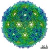
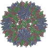
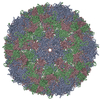
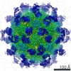
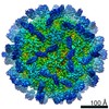
| Title | MS2 coat protein dimer with 145-GGGSYATMPIAKHVKDVGGGSGT-167 insertion VLP displaying fullerene C74-like D3 symmetry | |||||||||
 Map data Map data | main volume | |||||||||
 Sample Sample |
| |||||||||
| Function / homology | negative regulation of viral translation / Levivirus coat protein / Levivirus coat protein / Bacteriophage RNA-type, capsid / T=3 icosahedral viral capsid / structural molecule activity / RNA binding / identical protein binding / Capsid protein Function and homology information Function and homology information | |||||||||
| Biological species |  Escherichia phage MS2 (virus) / Escherichia virus MS2 Escherichia phage MS2 (virus) / Escherichia virus MS2 | |||||||||
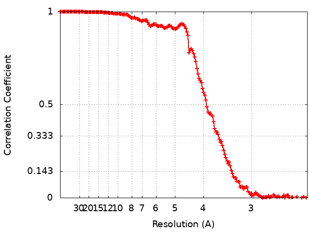
| Method | single particle reconstruction / cryo EM / Resolution: 3.34 Å | |||||||||
 Authors Authors | Biela AP | |||||||||
| Funding support |  Poland, 2 items Poland, 2 items
| |||||||||
 Citation Citation | Journal: Commun Mater / Year: 2022 Title: Programmable polymorphism of a virus-like particle. Authors: Artur P Biela / Antonina Naskalska / Farzad Fatehi / Reidun Twarock / Jonathan G Heddle /  Abstract: Virus-like particles (VLPs) have significant potential as artificial vaccines and drug delivery systems. The ability to control their size has wide ranging utility but achieving such controlled ...Virus-like particles (VLPs) have significant potential as artificial vaccines and drug delivery systems. The ability to control their size has wide ranging utility but achieving such controlled polymorphism using a single protein subunit is challenging as it requires altering VLP geometry. Here we achieve size control of MS2 bacteriophage VLPs via insertion of amino acid sequences in an external loop to shift morphology to significantly larger forms. The resulting VLP size and geometry is controlled by altering the length and type of the insert. Cryo electron microscopy structures of the new VLPs, in combination with a kinetic model of their assembly, show that the abundance of wild type ( = 3), = 4, D3 and D5 symmetrical VLPs can be biased in this way. We propose a mechanism whereby the insert leads to a change in the dynamic behavior of the capsid protein dimer, affecting the interconversion between the symmetric and asymmetric conformers and thus determining VLP size and morphology. | |||||||||
| History |
|
- Structure visualization
| Movie |
Movie viewer |
|---|---|
| Structure viewer | EM map: SurfViewMolmilJmol/JSmol |
| Supplemental images |

- Downloads & links
-EMDB archive
| Map data | emd_12788.map.gz | 328 MB | EMDB map data format | |
|---|---|---|---|---|
| Header (meta data) | emd-12788-v30.xmlemd-12788.xml | 11.2 KB 11.2 KB | Display Display | EMDB header |
| FSC (resolution estimation) | emd_12788_fsc.xml | 16.2 KB | Display | FSC data file |
| Images |  emd_12788.png emd_12788.png | 142.4 KB | ||
| Archive directory |  http://ftp.pdbj.org/pub/emdb/structures/EMD-12788ftp://ftp.pdbj.org/pub/emdb/structures/EMD-12788 http://ftp.pdbj.org/pub/emdb/structures/EMD-12788ftp://ftp.pdbj.org/pub/emdb/structures/EMD-12788 | HTTPS FTP |
-Related structure data
| Related structure data | C: citing same article ( |
|---|---|
| Similar structure data |


-Links
| EMDB pages | EMDB (EBI/PDBe) / EMDataResource |
|---|---|
| Related items in Molecule of the Month |

-Map
| File | Download / File: emd_12788.map.gz / Format: CCP4 / Size: 347.6 MB / Type: IMAGE STORED AS FLOATING POINT NUMBER (4 BYTES) | ||||||||||||||||||||||||||||||||||||||||||||||||||||||||||||
|---|---|---|---|---|---|---|---|---|---|---|---|---|---|---|---|---|---|---|---|---|---|---|---|---|---|---|---|---|---|---|---|---|---|---|---|---|---|---|---|---|---|---|---|---|---|---|---|---|---|---|---|---|---|---|---|---|---|---|---|---|---|
| Annotation | main volume | ||||||||||||||||||||||||||||||||||||||||||||||||||||||||||||
| Projections & slices | Image control
Images are generated by Spider. | ||||||||||||||||||||||||||||||||||||||||||||||||||||||||||||
| Voxel size | X=Y=Z: 1.14667 Å | ||||||||||||||||||||||||||||||||||||||||||||||||||||||||||||
| Density |
| ||||||||||||||||||||||||||||||||||||||||||||||||||||||||||||
| Symmetry | Space group: 1 | ||||||||||||||||||||||||||||||||||||||||||||||||||||||||||||
| Details | EMDB XML:
CCP4 map header:
| ||||||||||||||||||||||||||||||||||||||||||||||||||||||||||||
 Z (Sec.)
 Y (Row.)
 X (Col.)


-Supplemental data
- Sample components
-Entire : Escherichia virus MS2
| Entire | Name: Escherichia virus MS2 |
|---|---|
| Components |
|
-Supramolecule #1: Escherichia virus MS2
| Supramolecule | Name: Escherichia virus MS2 / type: virus / ID: 1 / Parent: 0 / Macromolecule list: all / NCBI-ID: 329852 / Sci species name: Escherichia virus MS2 / Virus type: VIRUS-LIKE PARTICLE / Virus isolate: OTHER / Virus enveloped: No / Virus empty: Yes |
|---|---|
| Host system | Organism:  |
| Molecular weight | Experimental: 3.3 MDa |
| Virus shell | Shell ID: 1 / Diameter: 340.0 Å |
-Macromolecule #1: MS2 bacteriophage coat protein dimer
| Macromolecule | Name: MS2 bacteriophage coat protein dimer / type: protein_or_peptide / ID: 1 Details: MS2 coat protein dimer with 145-GGGSYATMPIAKHVKDVGGGSGT-167 insertion Enantiomer: LEVO |
|---|---|
| Source (natural) | Organism: Escherichia phage MS2 (virus) |
| Recombinant expression | Organism: |
| Sequence | String: MASNFTPFVL VDNGGTGDVT VAPSNFANGV AEWISSNSRS QAYKVTCSVR QSSAQNRKYT IKVEVPKVAT QTVGGVELPV AAWRSYLNME LTIPIFATNS DCELIVKAMQ GLLKDGNPIP SAIAANSGIY ANFTQFVLVD NGGTGGGSYA TMPIAKHVKD VGGGSGTGDV ...String: MASNFTPFVL VDNGGTGDVT VAPSNFANGV AEWISSNSRS QAYKVTCSVR QSSAQNRKYT IKVEVPKVAT QTVGGVELPV AAWRSYLNME LTIPIFATNS DCELIVKAMQ GLLKDGNPIP SAIAANSGIY ANFTQFVLVD NGGTGGGSYA TMPIAKHVKD VGGGSGTGDV TVAPSNFANG VAEWISSNSR SQAYKVTCSV RQSSAQNRKY TIKVEVPKVA TQTVGGVELP VAAWRSYLNM ELTIPIFATN SDCELIVKAM QGLLKDGNPI PSAIAANSGI Y |
-Experimental details
-Structure determination
| Method | cryo EM |
|---|---|
 Processing Processing | single particle reconstruction |
| Aggregation state | particle |
-Sample preparation
| Concentration | 1 mg/mL |
|---|---|
| Buffer | pH: 7.4 |
| Grid | Model: Quantifoil R1.2/1.3 / Material: COPPER / Mesh: 400 / Pretreatment - Type: GLOW DISCHARGE / Pretreatment - Atmosphere: AIR / Pretreatment - Pressure: 101.325 kPa |
| Vitrification | Cryogen name: ETHANE / Chamber humidity: 100 % / Chamber temperature: 277 K / Instrument: FEI VITROBOT MARK IV |
- Electron microscopy
| Microscope | FEI TITAN KRIOS |
|---|---|
| Image recording | Film or detector model: GATAN K3 (6k x 4k) / Number grids imaged: 1 / Number real images: 4546 / Average electron dose: 40.0 e/Å2 |
| Electron beam | Acceleration voltage: 300 kV / Electron source:  FIELD EMISSION GUN FIELD EMISSION GUN |
| Electron optics | Illumination mode: FLOOD BEAM / Imaging mode: BRIGHT FIELD |
| Experimental equipment |  Model: Titan Krios / Image courtesy: FEI Company |