 ムービー
ムービー コントローラー


+ データを開く

- 基本情報
基本情報
| 登録情報 | データベース: EMDB / ID: EMD-1272 | |||||||||
|---|---|---|---|---|---|---|---|---|---|---|
| タイトル | Cryoelectron microscopy of protein IX-modified adenoviruses suggests a new position for the C terminus of protein IX. | |||||||||
 マップデータ マップデータ | This is the reconstruction of Ad-IX-GFP capsids. The data were collected at 2.17 angstrom per pixel and downsampled by 2 for processing. The final reconstruction was guassian lowpass filtered to 22 angstroms. The map deposited here is oriented with the 3-fold axis along z. This map is a half-map. | |||||||||
 試料 試料 |
| |||||||||
| 生物種 |   Human adenovirus 5 (ヒトアデノウイルス) Human adenovirus 5 (ヒトアデノウイルス) | |||||||||
| 手法 | 単粒子再構成法 / クライオ電子顕微鏡法 / 解像度: 22.0 Å | |||||||||
 データ登録者 データ登録者 | Marsh MP / Campos SK / Baker ML / Chen CY / Chiu W / Barry MA | |||||||||
 引用 引用 | ジャーナル: J Virol / 年: 2006 タイトル: Cryoelectron microscopy of protein IX-modified adenoviruses suggests a new position for the C terminus of protein IX. 著者: Michael P Marsh / Samuel K Campos / Matthew L Baker / Christopher Y Chen / Wah Chiu / Michael A Barry /  要旨: Recombinant human adenovirus is a useful gene delivery vector for clinical gene therapy. Minor capsid protein IX of adenovirus has been of recent interest since multiple studies have shown that ...Recombinant human adenovirus is a useful gene delivery vector for clinical gene therapy. Minor capsid protein IX of adenovirus has been of recent interest since multiple studies have shown that modifications can be made to its C terminus to alter viral tropism or add molecular tags and/or reporter proteins. We examined the structure of an engineered adenovirus displaying the enhanced green fluorescent protein (EGFP) fused to the C terminus of protein IX. Cryoelectron microscopy and reconstruction localized the C-terminal EGFP fusion between the H2 hexon and the H4 hexon, positioned between adjacent facets, directly above the density previously assigned as protein IIIa. The original assignment of IIIa was based largely on indirect evidence, and the data presented herein support the reassignment of the IIIa density as protein IX. | |||||||||
| 履歴 |
|
- 構造の表示
構造の表示
| ムービー |
 ムービービューア ムービービューア |
|---|---|
| 構造ビューア | EMマップ: SurfViewMolmilJmol/JSmol |
| 添付画像 |

- ダウンロードとリンク
-EMDBアーカイブ
| マップデータ | emd_1272.map.gz | 32.7 MB | EMDBマップデータ形式 | |
|---|---|---|---|---|
| ヘッダ (付随情報) | emd-1272-v30.xmlemd-1272.xml | 9.3 KB 9.3 KB | 表示 表示 | EMDBヘッダ |
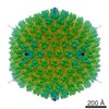
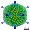
| 画像 | 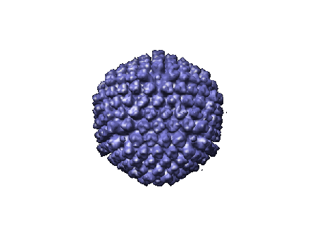 1272.gif 1272.gif | 27.1 KB | ||
| アーカイブディレクトリ |  http://ftp.pdbj.org/pub/emdb/structures/EMD-1272ftp://ftp.pdbj.org/pub/emdb/structures/EMD-1272 http://ftp.pdbj.org/pub/emdb/structures/EMD-1272ftp://ftp.pdbj.org/pub/emdb/structures/EMD-1272 | HTTPS FTP |
-検証レポート
| 文書・要旨 | emd_1272_validation.pdf.gz | 207.8 KB | 表示 | EMDB検証レポート |
|---|---|---|---|---|
| 文書・詳細版 | emd_1272_full_validation.pdf.gz | 207 KB | 表示 | |
| XML形式データ | emd_1272_validation.xml.gz | 4.9 KB | 表示 | |
| アーカイブディレクトリ | https://ftp.pdbj.org/pub/emdb/validation_reports/EMD-1272ftp://ftp.pdbj.org/pub/emdb/validation_reports/EMD-1272 | HTTPS FTP |
-関連構造データ
| 類似構造データ |
|---|
-リンク
| EMDBのページ | EMDB (EBI/PDBe) / EMDataResource |
|---|
-マップ
| ファイル | ダウンロード / ファイル: emd_1272.map.gz / 形式: CCP4 / 大きさ: 60.7 MB / タイプ: IMAGE STORED AS FLOATING POINT NUMBER (4 BYTES) | ||||||||||||||||||||||||||||||||||||||||||||||||||||||||||||||||||||
|---|---|---|---|---|---|---|---|---|---|---|---|---|---|---|---|---|---|---|---|---|---|---|---|---|---|---|---|---|---|---|---|---|---|---|---|---|---|---|---|---|---|---|---|---|---|---|---|---|---|---|---|---|---|---|---|---|---|---|---|---|---|---|---|---|---|---|---|---|---|
| 注釈 | This is the reconstruction of Ad-IX-GFP capsids. The data were collected at 2.17 angstrom per pixel and downsampled by 2 for processing. The final reconstruction was guassian lowpass filtered to 22 angstroms. The map deposited here is oriented with the 3-fold axis along z. This map is a half-map. | ||||||||||||||||||||||||||||||||||||||||||||||||||||||||||||||||||||
| 投影像・断面図 | 画像のコントロール
画像は Spider により作成 これらの図は立方格子座標系で作成されたものです | ||||||||||||||||||||||||||||||||||||||||||||||||||||||||||||||||||||
| ボクセルのサイズ | X=Y=Z: 4.34 Å | ||||||||||||||||||||||||||||||||||||||||||||||||||||||||||||||||||||
| 密度 |
| ||||||||||||||||||||||||||||||||||||||||||||||||||||||||||||||||||||
| 対称性 | 空間群: 1 | ||||||||||||||||||||||||||||||||||||||||||||||||||||||||||||||||||||
| 詳細 | EMDB XML:
CCP4マップ ヘッダ情報:
| ||||||||||||||||||||||||||||||||||||||||||||||||||||||||||||||||||||
 Z (Sec.)
 Y (Row.)
 X (Col.)


-添付データ
- 試料の構成要素
-全体 : Adenovirus capsid with GFP fused to C-terminus of capisd protein pIX
| 全体 | 名称: Adenovirus capsid with GFP fused to C-terminus of capisd protein pIX |
|---|---|
| 要素 |
|
-超分子 #1000: Adenovirus capsid with GFP fused to C-terminus of capisd protein pIX
| 超分子 | 名称: Adenovirus capsid with GFP fused to C-terminus of capisd protein pIX タイプ: sample / ID: 1000 / Number unique components: 1 |
|---|---|
| 分子量 | 理論値: 150 MDa |
-超分子 #1: Human adenovirus 5
| 超分子 | 名称: Human adenovirus 5 / タイプ: virus / ID: 1 / Name.synonym: Adenovirus, Ad-IX-GFP / 詳細: GFP fusion to protein pIX / NCBI-ID: 28285 / 生物種: Human adenovirus 5 / ウイルスタイプ: VIRION / ウイルス・単離状態: SEROTYPE / ウイルス・エンベロープ: No / ウイルス・中空状態: No / Syn species name: Adenovirus, Ad-IX-GFP |
|---|---|
| 宿主 | 生物種:  Homo sapiens (ヒト) / 別称: VERTEBRATES Homo sapiens (ヒト) / 別称: VERTEBRATES |
| 分子量 | 実験値: 150 MDa |
| ウイルス殻 | Shell ID: 1 / 名称: capsid / 直径: 920 Å / T番号(三角分割数): 25 |
-実験情報
-構造解析
| 手法 | クライオ電子顕微鏡法 |
|---|---|
 解析 解析 | 単粒子再構成法 |
| 試料の集合状態 | particle |
-試料調製
| 緩衝液 | pH: 7.2 / 詳細: Phosphate-buffered Saline (PBS) |
|---|---|
| グリッド | 詳細: 200 mesh copper grid |
| 凍結 | 凍結剤: ETHANE / チャンバー内湿度: 100 % / チャンバー内温度: 77 K / 装置: OTHER / 詳細: Vitrification instrument: Vitrobot / 手法: Blotted twice for one second each. |
- 電子顕微鏡法
| 顕微鏡 | JEOL 2010F |
|---|---|
| 温度 | 最低: 77 K |
| 日付 | 2004年12月17日 |
| 撮影 | カテゴリ: CCD フィルム・検出器のモデル: GATAN ULTRASCAN 4000 (4k x 4k) デジタル化 - サンプリング間隔: 2.17 µm / 実像数: 100 / 平均電子線量: 15 e/Å2 / ビット/ピクセル: 16 |
| 電子線 | 加速電圧: 200 kV / 電子線源:  FIELD EMISSION GUN FIELD EMISSION GUN |
| 電子光学系 | 照射モード: FLOOD BEAM / 撮影モード: BRIGHT FIELD / Cs: 2.0 mm / 最大 デフォーカス(公称値): 18.0 µm / 最小 デフォーカス(公称値): 0.5 µm / 倍率(公称値): 50000 |
| 試料ステージ | 試料ホルダー: Side entry liquid nitrogen-cooled cryo specimen holder.This holder operates in the temperature range from 77K to ambient. 試料ホルダーモデル: GATAN LIQUID NITROGEN |
-画像解析
| CTF補正 | 詳細: Each micrograph |
|---|---|
| 最終 再構成 | 想定した対称性 - 点群: I (正20面体型対称) / アルゴリズム: OTHER / 解像度のタイプ: BY AUTHOR / 解像度: 22.0 Å / 解像度の算出法: FSC 0.5 CUT-OFF / ソフトウェア - 名称: SAVR / 使用した粒子像数: 800 |
