+データを開く
-基本情報
登録情報 | データベース: EMDB / ID: EMD-1231 | |||||||||
---|---|---|---|---|---|---|---|---|---|---|
タイトル | Structure of the connector of bacteriophage T7 at 8A resolution: structural homologies of a basic component of a DNA translocating machinery. | |||||||||
マップデータ | 3D map of T7-connector | |||||||||
試料 |
| |||||||||
生物種 | Enterobacteria phage T7 (ファージ) | |||||||||
手法 | 単粒子再構成法 / クライオ電子顕微鏡法 / ネガティブ染色法 / 解像度: 8.0 Å | |||||||||
データ登録者 | Agirrezabala X / Martin-Benito J / Valle M / Gonzalez JM / Valencia A / Valpuesta JM / Carrascosa JL | |||||||||
引用 | 
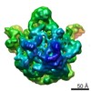
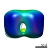
ジャーナル: J Mol Biol / 年: 2005 タイトル: Structure of the connector of bacteriophage T7 at 8A resolution: structural homologies of a basic component of a DNA translocating machinery. 著者: Xabier Agirrezabala / Jaime Martín-Benito / Mikel Valle / José M González / Alfonso Valencia / José María Valpuesta / José L Carrascosa / 要旨: The three-dimensional structure of the bacteriophage T7 head-to-tail connector has been obtained at 8A resolution using cryo-electron microscopy and single-particle analysis from purified recombinant ...The three-dimensional structure of the bacteriophage T7 head-to-tail connector has been obtained at 8A resolution using cryo-electron microscopy and single-particle analysis from purified recombinant connectors. The general morphology of the T7 connector is that of a 12-folded toroidal homopolymer with a channel that runs along the longitudinal axis of the particle. The structure of the T7 connector reveals many structural similarities with the connectors from other bacteriophages. Docking of the atomic structure of the varphi29 connector into the three-dimensional reconstruction of T7 connector reveals that the narrow, distal region of the two oligomers are almost identical. This region of the varphi29 connector has been suggested to be involved in DNA translocation, and is composed of an alpha-beta-alpha-beta-beta-alpha motif. A search for alpha-helices in the same region of the T7 three-dimensional map has located three alpha-helices in approximately the same position as those of the varphi29 connector. A comparison of the predicted secondary structure of several bacteriophage connectors, including among others T7, varphi29, P22 and SPP1, reveals that, despite the lack of sequence homology, they seem to contain the same alpha-beta-alpha-beta-beta-alpha motif as that present in the varphi29 connector. These results allow us to suggest a common architecture related to a basic component of the DNA translocating machinery for several viruses. | |||||||||
履歴 |
|
-構造の表示
ムービー |
ムービービューア |
---|---|
構造ビューア | EMマップ: SurfViewMolmilJmol/JSmol |
添付画像 |
-ダウンロードとリンク
-EMDBアーカイブ
マップデータ | emd_1231.map.gz | 9.6 MB | EMDBマップデータ形式 | |
---|---|---|---|---|
ヘッダ (付随情報) | emd-1231-v30.xml emd-1231.xml | 19.7 KB 19.7 KB | 表示 表示 | EMDBヘッダ |
画像 | 1231.gif | 38.6 KB | ||
アーカイブディレクトリ | http://ftp.pdbj.org/pub/emdb/structures/EMD-1231
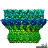
 ftp://ftp.pdbj.org/pub/emdb/structures/EMD-1231 | HTTPS FTP |
-関連構造データ
-リンク
EMDBのページ | EMDB (EBI/PDBe) / EMDataResource |
---|
-マップ
ファイル | ダウンロード / ファイル: emd_1231.map.gz / 形式: CCP4 / 大きさ: 10.2 MB / タイプ: IMAGE STORED AS FLOATING POINT NUMBER (4 BYTES) | ||||||||||||||||||||||||||||||||||||||||||||||||||||||||||||||||||||
---|---|---|---|---|---|---|---|---|---|---|---|---|---|---|---|---|---|---|---|---|---|---|---|---|---|---|---|---|---|---|---|---|---|---|---|---|---|---|---|---|---|---|---|---|---|---|---|---|---|---|---|---|---|---|---|---|---|---|---|---|---|---|---|---|---|---|---|---|---|
注釈 | 3D map of T7-connector | ||||||||||||||||||||||||||||||||||||||||||||||||||||||||||||||||||||
ボクセルのサイズ | X=Y=Z: 2.18 Å | ||||||||||||||||||||||||||||||||||||||||||||||||||||||||||||||||||||
密度 |
| ||||||||||||||||||||||||||||||||||||||||||||||||||||||||||||||||||||
対称性 | 空間群: 1 | ||||||||||||||||||||||||||||||||||||||||||||||||||||||||||||||||||||
詳細 | EMDB XML:
CCP4マップ ヘッダ情報:
|
-添付データ
-試料の構成要素
+全体 : Connector of Bacteriophage T7
+超分子 #1000: Connector of Bacteriophage T7
+分子 #1: gp8
+分子 #2: gp8
+分子 #3: gp8
+分子 #4: gp8
+分子 #5: gp8
+分子 #6: gp8
+分子 #7: gp8
+分子 #8: gp8
+分子 #9: gp8
+分子 #10: gp8
+分子 #11: gp8
+分子 #12: gp8
-実験情報
-構造解析
手法 | ネガティブ染色法, クライオ電子顕微鏡法 |
---|---|
解析 | 単粒子再構成法 |
試料の集合状態 | particle |
-試料調製
濃度 | 0.4 mg/mL |
---|---|
緩衝液 | pH: 7.8 詳細: 20mM Tris HCl pH:7.8, 3mM B-mercaptoethanol, 4%(v/v) glicerol, 2mM Na3EDTA, 100 mM NaCl |
染色 | タイプ: NEGATIVE / 詳細: cryoEM |
グリッド | 詳細: Quantifoil grids with 2 um hole size |
凍結 | 凍結剤: ETHANE |
-電子顕微鏡法
顕微鏡 | FEI TECNAI F20 |
---|---|
アライメント法 | Legacy - 非点収差: FFT with ssCCD |
撮影 | カテゴリ: FILM / フィルム・検出器のモデル: KODAK SO-163 FILM / デジタル化 - スキャナー: ZEISS SCAI / デジタル化 - サンプリング間隔: 14 µm / 平均電子線量: 10 e/Å2 |
Tilt angle min | 0 |
Tilt angle max | 0 |
電子線 | 加速電圧: 200 kV / 電子線源: FIELD EMISSION GUN |
電子光学系 | 倍率(補正後): 64220 / 照射モード: FLOOD BEAM / 撮影モード: BRIGHT FIELD / Cs: 2.26 mm / 最大 デフォーカス(公称値): 2.3 µm / 最小 デフォーカス(公称値): 0.9 µm / 倍率(公称値): 62000 |
試料ステージ | 試料ホルダー: Side entry liquid nitrogen-cooled cryo specimen holder, GATAN. Eucentric 試料ホルダーモデル: GATAN LIQUID NITROGEN |
実験機器 | モデル: Tecnai F20 / 画像提供: FEI Company |
-画像解析
CTF補正 | 詳細: Wiener filter, defocus groups |
---|---|
最終 再構成 | 想定した対称性 - 点群: C12 (12回回転対称) / アルゴリズム: OTHER / 解像度のタイプ: BY AUTHOR / 解像度: 8.0 Å / 解像度の算出法: FSC 0.5 CUT-OFF / ソフトウェア - 名称: SPIDER / 使用した粒子像数: 26492 |
最終 角度割当 | 詳細: SPIDER |