 Movie
Movie Controller

[English] 日本語
 Yorodumi
- EMDB-1209: Three-dimensional structure of the human DNA-PKcs/Ku70/Ku80 compl... -
+ Open data

- Basic information
| Entry | Database: EMDB / ID: EMD-1209 | |||||||||
|---|---|---|---|---|---|---|---|---|---|---|
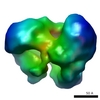
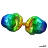
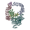
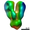
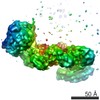
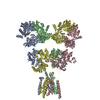
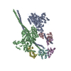
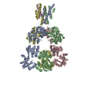
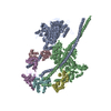
| Title | Three-dimensional structure of the human DNA-PKcs/Ku70/Ku80 complex assembled on DNA and its implications for DNA DSB repair. | |||||||||
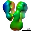
 Map data Map data | 3D reconstruction of the DNA-bound DNAPKcs-Ku70-Ku80 complex obtained from negatively stained samples | |||||||||
 Sample Sample |
| |||||||||
| Function / homology | : / Ku70 / Phosphatidylinositol 3-/4-kinase, catalytic domain / double-strand break repair via nonhomologous end joining / double-strand break repair / protein kinase activity Function and homology information Function and homology information | |||||||||
| Biological species |  Homo sapiens (human) Homo sapiens (human) | |||||||||
| Method | single particle reconstruction / negative staining / Resolution: 25.0 Å | |||||||||
 Authors Authors | Spagnolo L / Rivera-Calzada A / Pearl LH / Llorca O | |||||||||
 Citation Citation | Journal: Mol Cell / Year: 2006 Title: Three-dimensional structure of the human DNA-PKcs/Ku70/Ku80 complex assembled on DNA and its implications for DNA DSB repair. Authors: Laura Spagnolo / Angel Rivera-Calzada / Laurence H Pearl / Oscar Llorca /  Abstract: DNA-PKcs is a large (approximately 470 kDa) kinase that plays an essential role in the repair of DNA double-strand breaks (DSBs) by nonhomologous end joining (NHEJ). DNA-PKcs is recruited to DSBs by ...DNA-PKcs is a large (approximately 470 kDa) kinase that plays an essential role in the repair of DNA double-strand breaks (DSBs) by nonhomologous end joining (NHEJ). DNA-PKcs is recruited to DSBs by the Ku70/Ku80 heterodimer, with which it forms the core of a multiprotein complex that promotes synapsis of the broken DNA ends. We have purified the human DNA-PKcs/Ku70/Ku80 holoenzyme assembled on a DNA molecule. Its three-dimensional (3D) structure at approximately 25 Angstroms resolution was determined by single-particle electron microscopy. Binding of Ku and DNA elicits conformational changes in the FAT and FATC domains of DNA-PKcs. Dimeric particles are observed in which two DNA-PKcs/Ku70/Ku80 holoenzymes interact through the N-terminal HEAT repeats. The proximity of the dimer contacts to the likely positions of the DNA ends suggests that these represent synaptic complexes that maintain broken DNA ends in proximity and provide a platform for access of the various enzymes required for end processing and ligation. | |||||||||
| History |
|
- Structure visualization
| Movie |
Movie viewer |
|---|---|
| Structure viewer | EM map: SurfViewMolmilJmol/JSmol |
| Supplemental images |
 UCSF Chimera
- Downloads & links
-EMDB archive
| Map data | emd_1209.map.gz | 1.2 MB | EMDB map data format | |
|---|---|---|---|---|
| Header (meta data) | emd-1209-v30.xmlemd-1209.xml | 11.5 KB 11.5 KB | Display Display | EMDB header |
| Images |  1209.gif 1209.gif | 58.7 KB | ||
| Archive directory |  http://ftp.pdbj.org/pub/emdb/structures/EMD-1209ftp://ftp.pdbj.org/pub/emdb/structures/EMD-1209 http://ftp.pdbj.org/pub/emdb/structures/EMD-1209ftp://ftp.pdbj.org/pub/emdb/structures/EMD-1209 | HTTPS FTP |
-Validation report
| Summary document | emd_1209_validation.pdf.gz | 212.8 KB | Display | EMDB validaton report |
|---|---|---|---|---|
| Full document | emd_1209_full_validation.pdf.gz | 211.9 KB | Display | |
| Data in XML | emd_1209_validation.xml.gz | 5.2 KB | Display | |
| Arichive directory | https://ftp.pdbj.org/pub/emdb/validation_reports/EMD-1209ftp://ftp.pdbj.org/pub/emdb/validation_reports/EMD-1209 | HTTPS FTP |
-Related structure data

-Links
| EMDB pages | EMDB (EBI/PDBe) / EMDataResource |
|---|
-Map
| File | Download / File: emd_1209.map.gz / Format: CCP4 / Size: 1.4 MB / Type: IMAGE STORED AS FLOATING POINT NUMBER (4 BYTES) | ||||||||||||||||||||||||||||||||||||||||||||||||||||||||||||||||||||
|---|---|---|---|---|---|---|---|---|---|---|---|---|---|---|---|---|---|---|---|---|---|---|---|---|---|---|---|---|---|---|---|---|---|---|---|---|---|---|---|---|---|---|---|---|---|---|---|---|---|---|---|---|---|---|---|---|---|---|---|---|---|---|---|---|---|---|---|---|---|
| Annotation | 3D reconstruction of the DNA-bound DNAPKcs-Ku70-Ku80 complex obtained from negatively stained samples | ||||||||||||||||||||||||||||||||||||||||||||||||||||||||||||||||||||
| Projections & slices | Image control
Images are generated by Spider. | ||||||||||||||||||||||||||||||||||||||||||||||||||||||||||||||||||||
| Voxel size | X=Y=Z: 4.4 Å | ||||||||||||||||||||||||||||||||||||||||||||||||||||||||||||||||||||
| Density |
| ||||||||||||||||||||||||||||||||||||||||||||||||||||||||||||||||||||
| Symmetry | Space group: 1 | ||||||||||||||||||||||||||||||||||||||||||||||||||||||||||||||||||||
| Details | EMDB XML:
CCP4 map header:
| ||||||||||||||||||||||||||||||||||||||||||||||||||||||||||||||||||||
 Z (Sec.)
 Y (Row.)
 X (Col.)


-Supplemental data
- Sample components
-Entire : DNA-bound DNAPKcs-Ku70-Ku80 complex
| Entire | Name: DNA-bound DNAPKcs-Ku70-Ku80 complex |
|---|---|
| Components |
|
-Supramolecule #1000: DNA-bound DNAPKcs-Ku70-Ku80 complex
| Supramolecule | Name: DNA-bound DNAPKcs-Ku70-Ku80 complex / type: sample / ID: 1000 Oligomeric state: One DNA-PKcs molecule binds one Ku70 and one Ku80 on one DNA molecule Number unique components: 4 |
|---|---|
| Molecular weight | Theoretical: 650 KDa |
-Macromolecule #1: DNA-PKcs
| Macromolecule | Name: DNA-PKcs / type: protein_or_peptide / ID: 1 Name.synonym: DNA-dependent Protein Kinase catalytic subunit Number of copies: 1 / Oligomeric state: Monomer / Recombinant expression: Yes |
|---|---|
| Source (natural) | Organism: Homo sapiens (human) / synonym: Human / Cell: HeLa / Organelle: Nucleus / Location in cell: Nuclear |
| Molecular weight | Experimental: 470 KDa |
| Recombinant expression | Organism: HeLa cells |
| Sequence | GO: protein kinase activity InterPro: Phosphatidylinositol 3-/4-kinase, catalytic domain |
-Macromolecule #2: Ku70
| Macromolecule | Name: Ku70 / type: protein_or_peptide / ID: 2 / Number of copies: 1 / Oligomeric state: Monomer / Recombinant expression: Yes |
|---|---|
| Source (natural) | Organism: Homo sapiens (human) / synonym: Human / Cell: HeLa / Organelle: Nucleus / Location in cell: Nuclear |
| Molecular weight | Experimental: 70 KDa |
| Recombinant expression | Organism: HeLa cells |
| Sequence | GO: double-strand break repair via nonhomologous end joining InterPro: Ku70 |
-Macromolecule #3: Ku80
| Macromolecule | Name: Ku80 / type: protein_or_peptide / ID: 3 / Number of copies: 1 / Oligomeric state: Monomer / Recombinant expression: Yes |
|---|---|
| Source (natural) | Organism: Homo sapiens (human) / synonym: Human / Cell: HeLa / Organelle: Nucleus / Location in cell: Nuclear |
| Molecular weight | Experimental: 80 KDa |
| Recombinant expression | Organism: HeLa cells |
| Sequence | GO: double-strand break repair / InterPro: INTERPRO: IPR011210 |
-Macromolecule #4: DNA
| Macromolecule | Name: DNA / type: dna / ID: 4 / Classification: DNA / Structure: DOUBLE HELIX / Synthetic?: Yes |
|---|---|
| Sequence | String: ACGCGTGCGG CCATAATAAT AGTTTTTAGT TTATTGGGCG CG |
-Experimental details
-Structure determination
| Method | negative staining |
|---|---|
 Processing Processing | single particle reconstruction |
| Aggregation state | particle |
-Sample preparation
| Buffer | pH: 7.5 Details: 20 mM HEPES pH 7.5, 0.5 mM DTT, 0.25 mM EDTA, 0.0005% beta-octylglucoside, 25% glycerol, 100 mM NaCl |
|---|---|
| Staining | Type: NEGATIVE Details: The sample was applied to carbon-coated grids after glow-discharge and negatively stained with 1% uranyl acetate |
| Grid | Details: 400 mesh Rhodium-copper grids |
| Vitrification | Cryogen name: NONE |
- Electron microscopy
| Microscope | JEOL 1230 |
|---|---|
| Alignment procedure | Legacy - Astigmatism: correction with FFT and CCD camera |
| Details | Microscope: JEOL 1230 operated at 100 Kv Specimen holder, JEOL type M: 207EM-11020. |
| Image recording | Category: FILM / Film or detector model: KODAK 4489 FILM / Digitization - Scanner: OTHER / Digitization - Sampling interval: 10 µm / Number real images: 84 / Details: Scanner: MINOLTA Dimage Scan Multi Pro scanner / Bits/pixel: 16 |
| Tilt angle min | 0 |
| Tilt angle max | 0 |
| Electron beam | Acceleration voltage: 100 kV / Electron source: TUNGSTEN HAIRPIN |
| Electron optics | Illumination mode: FLOOD BEAM / Imaging mode: BRIGHT FIELD / Cs: 2.9 mm / Nominal magnification: 50000 |
| Sample stage | Specimen holder: JEOL type M / Specimen holder model: OTHER |
-Image processing
| Final reconstruction | Applied symmetry - Point group: C1 (asymmetric) / Algorithm: OTHER / Resolution.type: BY AUTHOR / Resolution: 25.0 Å / Resolution method: FSC 0.5 CUT-OFF / Software - Name: EMAN / Number images used: 14239 |
|---|
