 ムービー
ムービー コントローラー


+ データを開く

- 基本情報
基本情報
| 登録情報 | データベース: EMDB / ID: EMD-12047 | ||||||||||||
|---|---|---|---|---|---|---|---|---|---|---|---|---|---|
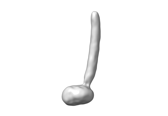
| タイトル | CryoEM Structure of the yeast peroxisomal membrane Pex14p/Pex17p complex | ||||||||||||
 マップデータ マップデータ | CryoEM Structure of the Pex14p/Pex17p complex | ||||||||||||
 試料 試料 |
| ||||||||||||
| 生物種 |  | ||||||||||||
| 手法 | 単粒子再構成法 / クライオ電子顕微鏡法 / 解像度: 10.2 Å | ||||||||||||
 データ登録者 データ登録者 | Lill P / Gatsogiannis C | ||||||||||||
| 資金援助 |  ドイツ, 3件 ドイツ, 3件
| ||||||||||||
 引用 引用 | ジャーナル: Proc Natl Acad Sci U S A / 年: 2020 タイトル: Towards the molecular architecture of the peroxisomal receptor docking complex. 著者: Pascal Lill / Tobias Hansen / Daniel Wendscheck / Bjoern Udo Klink / Tomasz Jeziorek / Dimitrios Vismpas / Jonas Miehling / Julian Bender / Andreas Schummer / Friedel Drepper / Wolfgang ...著者: Pascal Lill / Tobias Hansen / Daniel Wendscheck / Bjoern Udo Klink / Tomasz Jeziorek / Dimitrios Vismpas / Jonas Miehling / Julian Bender / Andreas Schummer / Friedel Drepper / Wolfgang Girzalsky / Bettina Warscheid / Ralf Erdmann / Christos Gatsogiannis / 要旨: Import of yeast peroxisomal matrix proteins is initiated by cytosolic receptors, which specifically recognize and bind the respective cargo proteins. At the peroxisomal membrane, the cargo-loaded ...Import of yeast peroxisomal matrix proteins is initiated by cytosolic receptors, which specifically recognize and bind the respective cargo proteins. At the peroxisomal membrane, the cargo-loaded receptor interacts with the docking protein Pex14p that is tightly associated with Pex17p. Previous data suggest that this interaction triggers the formation of an import pore for further translocation of the cargo. The mechanistic principles, however, are unclear, mainly because structures of higher-order assemblies are still lacking. Here, using an integrative approach, we provide the structural characterization of the major components of the peroxisomal docking complex Pex14p/Pex17p, in a native bilayer environment, and reveal its subunit organization. Our data show that three copies of Pex14p and a single copy of Pex17p assemble to form a 20-nm rod-like particle. The different subunits are arranged in a parallel manner, showing interactions along their complete sequences and providing receptor binding sites on both membrane sides. The long rod facing the cytosol is mainly formed by the predicted coiled-coil domains of Pex14p and Pex17p, possibly providing the necessary structural support for the formation of the import pore. Further implications of Pex14p/Pex17p for formation of the peroxisomal translocon are discussed. | ||||||||||||
| 履歴 |
|
- 構造の表示
構造の表示
| ムービー |
 ムービービューア ムービービューア |
|---|---|
| 構造ビューア | EMマップ: SurfViewMolmilJmol/JSmol |
| 添付画像 |
- ダウンロードとリンク
-EMDBアーカイブ
| マップデータ | emd_12047.map.gz | 6.3 MB | EMDBマップデータ形式 | |
|---|---|---|---|---|
| ヘッダ (付随情報) | emd-12047-v30.xmlemd-12047.xml | 17 KB 17 KB | 表示 表示 | EMDBヘッダ |
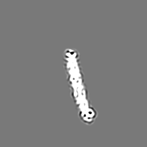
| 画像 |  emd_12047.png emd_12047.png | 21.5 KB | ||
| その他 | emd_12047_additional_1.map.gz | 3.6 MB | ||
| アーカイブディレクトリ |  http://ftp.pdbj.org/pub/emdb/structures/EMD-12047ftp://ftp.pdbj.org/pub/emdb/structures/EMD-12047 http://ftp.pdbj.org/pub/emdb/structures/EMD-12047ftp://ftp.pdbj.org/pub/emdb/structures/EMD-12047 | HTTPS FTP |
-検証レポート
| 文書・要旨 | emd_12047_validation.pdf.gz | 285.5 KB | 表示 | EMDB検証レポート |
|---|---|---|---|---|
| 文書・詳細版 | emd_12047_full_validation.pdf.gz | 285.1 KB | 表示 | |
| XML形式データ | emd_12047_validation.xml.gz | 6.4 KB | 表示 | |
| CIF形式データ | emd_12047_validation.cif.gz | 7.3 KB | 表示 | |
| アーカイブディレクトリ | https://ftp.pdbj.org/pub/emdb/validation_reports/EMD-12047ftp://ftp.pdbj.org/pub/emdb/validation_reports/EMD-12047 | HTTPS FTP |
-関連構造データ


-リンク
| EMDBのページ | EMDB (EBI/PDBe) / EMDataResource |
|---|
-マップ
| ファイル | ダウンロード / ファイル: emd_12047.map.gz / 形式: CCP4 / 大きさ: 103 MB / タイプ: IMAGE STORED AS FLOATING POINT NUMBER (4 BYTES) | ||||||||||||||||||||||||||||||||||||||||||||||||||||||||||||
|---|---|---|---|---|---|---|---|---|---|---|---|---|---|---|---|---|---|---|---|---|---|---|---|---|---|---|---|---|---|---|---|---|---|---|---|---|---|---|---|---|---|---|---|---|---|---|---|---|---|---|---|---|---|---|---|---|---|---|---|---|---|
| 注釈 | CryoEM Structure of the Pex14p/Pex17p complex | ||||||||||||||||||||||||||||||||||||||||||||||||||||||||||||
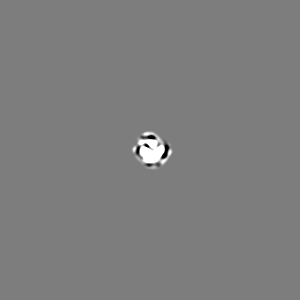
| 投影像・断面図 | 画像のコントロール
画像は Spider により作成 | ||||||||||||||||||||||||||||||||||||||||||||||||||||||||||||
| ボクセルのサイズ | X=Y=Z: 1.09 Å | ||||||||||||||||||||||||||||||||||||||||||||||||||||||||||||
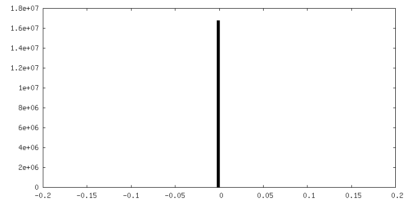
| 密度 |
| ||||||||||||||||||||||||||||||||||||||||||||||||||||||||||||
| 対称性 | 空間群: 1 | ||||||||||||||||||||||||||||||||||||||||||||||||||||||||||||
| 詳細 | EMDB XML:
CCP4マップ ヘッダ情報:
| ||||||||||||||||||||||||||||||||||||||||||||||||||||||||||||
 Z (Sec.)
 Y (Row.)
 X (Col.)


-添付データ
-追加マップ: CryoEM structure of the rod domain of the...
| ファイル | emd_12047_additional_1.map | ||||||||||||
|---|---|---|---|---|---|---|---|---|---|---|---|---|---|
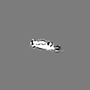
| 注釈 | CryoEM structure of the rod domain of the Pex14p/Pex17p complex upon signal subtraction of the nanodisc density and subsequent refinement. | ||||||||||||
| 投影像・断面図 |
| ||||||||||||
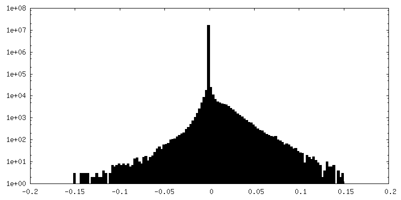
| 密度ヒストグラム |
- 試料の構成要素
-全体 : Peroxisomal Membrane Complex Pex14p/Pex17p reconstituted in cMSP1...
| 全体 | 名称: Peroxisomal Membrane Complex Pex14p/Pex17p reconstituted in cMSP1D1dH4-6 nanodiscs |
|---|---|
| 要素 |
|
-超分子 #1: Peroxisomal Membrane Complex Pex14p/Pex17p reconstituted in cMSP1...
| 超分子 | 名称: Peroxisomal Membrane Complex Pex14p/Pex17p reconstituted in cMSP1D1dH4-6 nanodiscs タイプ: complex / ID: 1 / 親要素: 0 / 含まれる分子: all 詳細: Pex14p and Pex17p constitute the major components of the peroxisomal membrane docking complex. Pex14p and Pex17p form a 3:1 heterotetrameric complex. |
|---|---|
| 由来(天然) | 生物種: |
| 分子量 | 理論値: 144 KDa |
-分子 #1: Peroxisomal membrane protein Pex14p
| 分子 | 名称: Peroxisomal membrane protein Pex14p / タイプ: protein_or_peptide / ID: 1 / 光学異性体: LEVO |
|---|---|
| 由来(天然) | 生物種: |
| 組換発現 | 生物種:  |
| 配列 | 文字列: MSDVVSKDRK ALFDSAVSFL KDESIKDAPL LKKIEFLKSK GLTEKEIEIA MKEPKKDGIV GDEVSKKIGS TENRASQDM YLYEAMPPTL PHRDWKDYFV MATATAGLLY GAYEVTRRYV IPNILPEAKS KLEGDKKEID D QFSKIDTV LNAIEAEQAE FRKKESETLK ...文字列: MSDVVSKDRK ALFDSAVSFL KDESIKDAPL LKKIEFLKSK GLTEKEIEIA MKEPKKDGIV GDEVSKKIGS TENRASQDM YLYEAMPPTL PHRDWKDYFV MATATAGLLY GAYEVTRRYV IPNILPEAKS KLEGDKKEID D QFSKIDTV LNAIEAEQAE FRKKESETLK ELSDTIAELK QALVQTTRSR EKIEDEFRIV KLEVVNMQNT ID KFVSDND GMQELNNIQK EMESLKSLMN NRMESGNAQD NRLFSISPNG IPGIDTIPSA SEILAKMGMQ EES DKEKEN GSDANKDDNA VPAWKKAREQ TIDSNASIPE WQKNTAANEI SVPDWQNGQV EDSIP |
-分子 #2: Peroxisomal membrane protein Pex17p
| 分子 | 名称: Peroxisomal membrane protein Pex17p / タイプ: protein_or_peptide / ID: 2 / 光学異性体: LEVO |
|---|---|
| 由来(天然) | 生物種: |
| 組換発現 | 生物種: |
| 配列 | 文字列: MTSINSFPRN IDWPSNIGIK KIEGTNPTVN AIKGLLYNGG SIYAFLYFVI AMFVEPTLQK QYQQRNDFSL FVLLRLRRI IAQLQKRLVM TPVSSLGFNE QNNFVERSTQ TSDDNIIRED NSHWAEMIYQ LQNMKQELQY F NRSSGQPS ESIDDFVFQI KMVTDQVELT ...文字列: MTSINSFPRN IDWPSNIGIK KIEGTNPTVN AIKGLLYNGG SIYAFLYFVI AMFVEPTLQK QYQQRNDFSL FVLLRLRRI IAQLQKRLVM TPVSSLGFNE QNNFVERSTQ TSDDNIIRED NSHWAEMIYQ LQNMKQELQY F NRSSGQPS ESIDDFVFQI KMVTDQVELT DRSRAFSNKS RNIIQGIREI KGWFVNGQVP R |
-実験情報
-構造解析
| 手法 | クライオ電子顕微鏡法 |
|---|---|
 解析 解析 | 単粒子再構成法 |
| 試料の集合状態 | particle |
-試料調製
| 濃度 | 0.15 mg/mL | ||||||
|---|---|---|---|---|---|---|---|
| 緩衝液 | pH: 8 / 構成要素:
| ||||||
| グリッド | モデル: Quantifoil / 材質: COPPER / メッシュ: 300 / 支持フィルム - 材質: CARBON / 支持フィルム - トポロジー: HOLEY / 前処理 - タイプ: GLOW DISCHARGE | ||||||
| 凍結 | 凍結剤: ETHANE / チャンバー内湿度: 100 % / チャンバー内温度: 277 K / 装置: FEI VITROBOT MARK II | ||||||
| 詳細 | The complex was reconstituted in circularMSP1D1D4-6 nanodiscs. |
- 電子顕微鏡法
| 顕微鏡 | FEI TITAN KRIOS |
|---|---|
| 特殊光学系 | 位相板: VOLTA PHASE PLATE |
| 撮影 | フィルム・検出器のモデル: GATAN K2 SUMMIT (4k x 4k) 検出モード: COUNTING / デジタル化 - 画像ごとのフレーム数: 1-40 / 撮影したグリッド数: 1 / 実像数: 3282 / 平均露光時間: 15.0 sec. / 平均電子線量: 56.0 e/Å2 |
| 電子線 | 加速電圧: 300 kV / 電子線源:  FIELD EMISSION GUN FIELD EMISSION GUN |
| 電子光学系 | 照射モード: FLOOD BEAM / 撮影モード: BRIGHT FIELD / Cs: 0.0 mm / 倍率(公称値): 130000 |
| 試料ステージ | 試料ホルダーモデル: FEI TITAN KRIOS AUTOGRID HOLDER ホルダー冷却材: NITROGEN |
| 実験機器 |  モデル: Titan Krios / 画像提供: FEI Company |
