 ムービー
ムービー コントローラー


+ データを開く

- 基本情報
基本情報
| 登録情報 | データベース: EMDB / ID: EMD-11967 | |||||||||
|---|---|---|---|---|---|---|---|---|---|---|
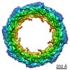
| タイトル | Human nuclear pore complex in HIV-1 infected T cells | |||||||||
 マップデータ マップデータ | Human Nuclear Pore Complex from HIV-1 infected T cells | |||||||||
 試料 試料 |
| |||||||||
| 生物種 |  Homo sapiens (ヒト) Homo sapiens (ヒト) | |||||||||
| 手法 | サブトモグラム平均法 / クライオ電子顕微鏡法 / 解像度: 37.0 Å | |||||||||
 データ登録者 データ登録者 | Margiotta E / Beck M | |||||||||
| 資金援助 |  ドイツ, 1件 ドイツ, 1件
| |||||||||
 引用 引用 | ジャーナル: Cell / 年: 2021 タイトル: Cone-shaped HIV-1 capsids are transported through intact nuclear pores. 著者: Vojtech Zila / Erica Margiotta / Beata Turoňová / Thorsten G Müller / Christian E Zimmerli / Simone Mattei / Matteo Allegretti / Kathleen Börner / Jona Rada / Barbara Müller / Marina ...著者: Vojtech Zila / Erica Margiotta / Beata Turoňová / Thorsten G Müller / Christian E Zimmerli / Simone Mattei / Matteo Allegretti / Kathleen Börner / Jona Rada / Barbara Müller / Marina Lusic / Hans-Georg Kräusslich / Martin Beck / 要旨: Human immunodeficiency virus (HIV-1) remains a major health threat. Viral capsid uncoating and nuclear import of the viral genome are critical for productive infection. The size of the HIV-1 capsid ...Human immunodeficiency virus (HIV-1) remains a major health threat. Viral capsid uncoating and nuclear import of the viral genome are critical for productive infection. The size of the HIV-1 capsid is generally believed to exceed the diameter of the nuclear pore complex (NPC), indicating that capsid uncoating has to occur prior to nuclear import. Here, we combined correlative light and electron microscopy with subtomogram averaging to capture the structural status of reverse transcription-competent HIV-1 complexes in infected T cells. We demonstrated that the diameter of the NPC in cellulo is sufficient for the import of apparently intact, cone-shaped capsids. Subsequent to nuclear import, we detected disrupted and empty capsid fragments, indicating that uncoating of the replication complex occurs by breaking the capsid open, and not by disassembly into individual subunits. Our data directly visualize a key step in HIV-1 replication and enhance our mechanistic understanding of the viral life cycle. #1: ジャーナル: To Be Publishedタイトル: Cone-shaped HIV-1 capsids are transported through intact nuclear pores 著者: Zila V / Margiotta E | |||||||||
| 履歴 |
|
- 構造の表示
構造の表示
| ムービー |
 ムービービューア ムービービューア |
|---|---|
| 構造ビューア | EMマップ: SurfViewMolmilJmol/JSmol |
| 添付画像 |

- ダウンロードとリンク
-EMDBアーカイブ
| マップデータ | emd_11967.map.gz | 2.5 MB | EMDBマップデータ形式 | |
|---|---|---|---|---|
| ヘッダ (付随情報) | emd-11967-v30.xmlemd-11967.xml | 9.4 KB 9.4 KB | 表示 表示 | EMDBヘッダ |
| 画像 | 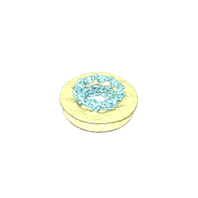 emd_11967.png emd_11967.png | 40.5 KB | ||
| アーカイブディレクトリ |  http://ftp.pdbj.org/pub/emdb/structures/EMD-11967ftp://ftp.pdbj.org/pub/emdb/structures/EMD-11967 http://ftp.pdbj.org/pub/emdb/structures/EMD-11967ftp://ftp.pdbj.org/pub/emdb/structures/EMD-11967 | HTTPS FTP |
-検証レポート
| 文書・要旨 | emd_11967_validation.pdf.gz | 214.5 KB | 表示 | EMDB検証レポート |
|---|---|---|---|---|
| 文書・詳細版 | emd_11967_full_validation.pdf.gz | 213.6 KB | 表示 | |
| XML形式データ | emd_11967_validation.xml.gz | 5.7 KB | 表示 | |
| アーカイブディレクトリ | https://ftp.pdbj.org/pub/emdb/validation_reports/EMD-11967ftp://ftp.pdbj.org/pub/emdb/validation_reports/EMD-11967 | HTTPS FTP |
-関連構造データ
-リンク
| EMDBのページ | EMDB (EBI/PDBe) / EMDataResource |
|---|
-マップ
| ファイル | ダウンロード / ファイル: emd_11967.map.gz / 形式: CCP4 / 大きさ: 11.4 MB / タイプ: IMAGE STORED AS FLOATING POINT NUMBER (4 BYTES) | ||||||||||||||||||||||||||||||||||||||||||||||||||||||||||||
|---|---|---|---|---|---|---|---|---|---|---|---|---|---|---|---|---|---|---|---|---|---|---|---|---|---|---|---|---|---|---|---|---|---|---|---|---|---|---|---|---|---|---|---|---|---|---|---|---|---|---|---|---|---|---|---|---|---|---|---|---|---|
| 注釈 | Human Nuclear Pore Complex from HIV-1 infected T cells | ||||||||||||||||||||||||||||||||||||||||||||||||||||||||||||
| 投影像・断面図 | 画像のコントロール
画像は Spider により作成 | ||||||||||||||||||||||||||||||||||||||||||||||||||||||||||||
| ボクセルのサイズ | X=Y=Z: 13.8 Å | ||||||||||||||||||||||||||||||||||||||||||||||||||||||||||||
| 密度 |
| ||||||||||||||||||||||||||||||||||||||||||||||||||||||||||||
| 対称性 | 空間群: 1 | ||||||||||||||||||||||||||||||||||||||||||||||||||||||||||||
| 詳細 | EMDB XML:
CCP4マップ ヘッダ情報:
| ||||||||||||||||||||||||||||||||||||||||||||||||||||||||||||
 Z (Sec.)
 Y (Row.)
 X (Col.)


-添付データ
- 試料の構成要素
-全体 : Nuclear pore complex from HIV-1 infected T cells
| 全体 | 名称: Nuclear pore complex from HIV-1 infected T cells |
|---|---|
| 要素 |
|
-超分子 #1: Nuclear pore complex from HIV-1 infected T cells
| 超分子 | 名称: Nuclear pore complex from HIV-1 infected T cells / タイプ: complex / ID: 1 / 親要素: 0 |
|---|---|
| 由来(天然) | 生物種: Homo sapiens (ヒト) |
-実験情報
-構造解析
| 手法 | クライオ電子顕微鏡法 |
|---|---|
 解析 解析 | サブトモグラム平均法 |
| 試料の集合状態 | cell |
-試料調製
| 緩衝液 | pH: 7.4 / 詳細: RPMI 1640 medium with GlutaMAX |
|---|---|
| グリッド | モデル: Quantifoil / 材質: GOLD / メッシュ: 200 / 前処理 - タイプ: GLOW DISCHARGE |
| 凍結 | 凍結剤: ETHANE / チャンバー内湿度: 100 % / チャンバー内温度: 310.15 K / 装置: LEICA EM GP |
- 電子顕微鏡法
| 顕微鏡 | FEI TITAN KRIOS |
|---|---|
| 撮影 | フィルム・検出器のモデル: GATAN K2 QUANTUM (4k x 4k) 平均電子線量: 4.0 e/Å2 |
| 電子線 | 加速電圧: 300 kV / 電子線源:  FIELD EMISSION GUN FIELD EMISSION GUN |
| 電子光学系 | 照射モード: OTHER / 撮影モード: OTHER / 最大 デフォーカス(公称値): 5.0 µm / 最小 デフォーカス(公称値): 2.0 µm / 倍率(公称値): 42000 |
| 実験機器 |  モデル: Titan Krios / 画像提供: FEI Company |
-画像解析
| 最終 再構成 | 想定した対称性 - 点群: C8 (8回回転対称) / 解像度のタイプ: BY AUTHOR / 解像度: 37.0 Å / 解像度の算出法: FSC 0.143 CUT-OFF / 使用したサブトモグラム数: 90 |
|---|---|
| 抽出 | トモグラム数: 250 / 使用した粒子像数: 99 |
| CTF補正 | 詳細: novaCTF (Turonova et al., 2017) |
| 最終 角度割当 | タイプ: PROJECTION MATCHING |
