Movie
Movie Controller

[English] 日本語
 Yorodumi
- EMDB-11931: Subtomogram average of outer membrane dome protein (OMDP)-C8 symm... -
+ Open data

- Basic information
| Entry | Database: EMDB / ID: EMD-11931 | |||||||||
|---|---|---|---|---|---|---|---|---|---|---|
| Title | Subtomogram average of outer membrane dome protein (OMDP)-C8 symmetrized | |||||||||
 Map data Map data | STA of Outer membrane protein complexed-C8 symmetrized | |||||||||
 Sample Sample |
| |||||||||
| Biological species |  Homo sapiens (human) Homo sapiens (human) | |||||||||
| Method | subtomogram averaging / cryo EM / Resolution: 40.2 Å | |||||||||
 Authors Authors | Klein S / Wimmer B / Winter S / Kolovou A / Laketa V / Chlanda P | |||||||||
| Funding support |  Germany, 1 items Germany, 1 items
| |||||||||
 Citation Citation | Journal: Commun Biol / Year: 2021 Title: Post-correlation on-lamella cryo-CLEM reveals the membrane architecture of lamellar bodies. Authors: Steffen Klein / Benedikt H Wimmer / Sophie L Winter / Androniki Kolovou / Vibor Laketa / Petr Chlanda / Abstract: Lamellar bodies (LBs) are surfactant-rich organelles in alveolar cells. LBs disassemble into a lipid-protein network that reduces surface tension and facilitates gas exchange in the alveolar cavity. ...Lamellar bodies (LBs) are surfactant-rich organelles in alveolar cells. LBs disassemble into a lipid-protein network that reduces surface tension and facilitates gas exchange in the alveolar cavity. Current knowledge of LB architecture is predominantly based on electron microscopy studies using disruptive sample preparation methods. We established and validated a post-correlation on-lamella cryo-correlative light and electron microscopy approach for cryo-FIB milled cells to structurally characterize and validate the identity of LBs in their unperturbed state. Using deconvolution and 3D image registration, we were able to identify fluorescently labeled membrane structures analyzed by cryo-electron tomography. In situ cryo-electron tomography of A549 cells as well as primary Human Small Airway Epithelial Cells revealed that LBs are composed of membrane sheets frequently attached to the limiting membrane through "T"-junctions. We report a so far undescribed outer membrane dome protein complex (OMDP) on the limiting membrane of LBs. Our data suggest that LB biogenesis is driven by parallel membrane sheet import and by the curvature of the limiting membrane to maximize lipid storage capacity. | |||||||||
| History |
|
- Structure visualization
| Movie |
 Movie viewer Movie viewer |
|---|---|
| Structure viewer | EM map: SurfViewMolmilJmol/JSmol |
| Supplemental images |

- Downloads & links
-EMDB archive
| Map data | emd_11931.map.gz | 3.4 MB | EMDB map data format | |
|---|---|---|---|---|
| Header (meta data) | emd-11931-v30.xmlemd-11931.xml | 15.9 KB 15.9 KB | Display Display | EMDB header |
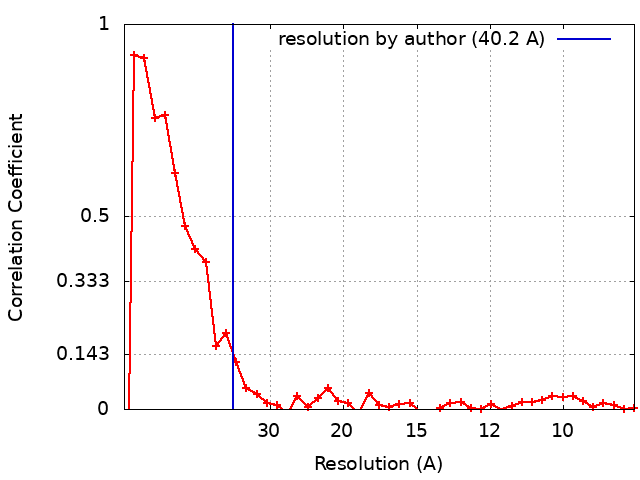
| FSC (resolution estimation) | emd_11931_fsc.xml | 4.7 KB | Display | FSC data file |
| Images |  emd_11931.png emd_11931.png | 185.7 KB | ||
| Others | emd_11931_half_map_1.map.gzemd_11931_half_map_2.map.gz | 3.5 MB 3.5 MB | ||
| Archive directory |  http://ftp.pdbj.org/pub/emdb/structures/EMD-11931ftp://ftp.pdbj.org/pub/emdb/structures/EMD-11931 http://ftp.pdbj.org/pub/emdb/structures/EMD-11931ftp://ftp.pdbj.org/pub/emdb/structures/EMD-11931 | HTTPS FTP |
-Related structure data


-Links
| EMDB pages | EMDB (EBI/PDBe) / EMDataResource |
|---|
-Map
| File | Download / File: emd_11931.map.gz / Format: CCP4 / Size: 3.8 MB / Type: IMAGE STORED AS FLOATING POINT NUMBER (4 BYTES) | ||||||||||||||||||||||||||||||||||||||||||||||||||||||||||||
|---|---|---|---|---|---|---|---|---|---|---|---|---|---|---|---|---|---|---|---|---|---|---|---|---|---|---|---|---|---|---|---|---|---|---|---|---|---|---|---|---|---|---|---|---|---|---|---|---|---|---|---|---|---|---|---|---|---|---|---|---|---|
| Annotation | STA of Outer membrane protein complexed-C8 symmetrized | ||||||||||||||||||||||||||||||||||||||||||||||||||||||||||||
| Projections & slices | Image control
Images are generated by Spider. | ||||||||||||||||||||||||||||||||||||||||||||||||||||||||||||
| Voxel size | X=Y=Z: 4.302 Å | ||||||||||||||||||||||||||||||||||||||||||||||||||||||||||||
| Density |
| ||||||||||||||||||||||||||||||||||||||||||||||||||||||||||||
| Symmetry | Space group: 1 | ||||||||||||||||||||||||||||||||||||||||||||||||||||||||||||
| Details | EMDB XML:
CCP4 map header:
| ||||||||||||||||||||||||||||||||||||||||||||||||||||||||||||
 Z (Sec.)
 Y (Row.)
 X (Col.)


-Supplemental data
-Half map: odd map
| File | emd_11931_half_map_1.map | ||||||||||||
|---|---|---|---|---|---|---|---|---|---|---|---|---|---|
| Annotation | odd map | ||||||||||||
| Projections & Slices |
| ||||||||||||
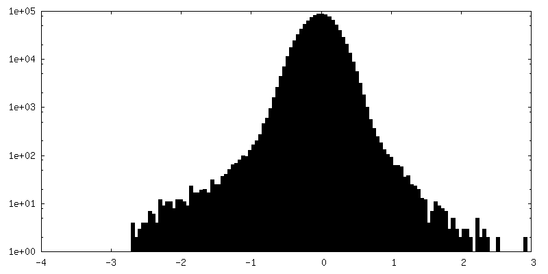
| Density Histograms |


-Half map: even map
| File | emd_11931_half_map_2.map | ||||||||||||
|---|---|---|---|---|---|---|---|---|---|---|---|---|---|
| Annotation | even map | ||||||||||||
| Projections & Slices |
| ||||||||||||
| Density Histograms |


- Sample components
-Entire : Subtomogram average of outer membrane dome protein (OMDP)-C8 symm...
| Entire | Name: Subtomogram average of outer membrane dome protein (OMDP)-C8 symmetrized |
|---|---|
| Components |
|
-Supramolecule #1: Subtomogram average of outer membrane dome protein (OMDP)-C8 symm...
| Supramolecule | Name: Subtomogram average of outer membrane dome protein (OMDP)-C8 symmetrized type: organelle_or_cellular_component / ID: 1 / Parent: 0 / Macromolecule list: #1 / Details: A549 human lung cell line |
|---|---|
| Source (natural) | Organism: Homo sapiens (human) |
-Experimental details
-Structure determination
| Method | cryo EM |
|---|---|
 Processing Processing | subtomogram averaging |
| Aggregation state | cell |
-Sample preparation
| Buffer | pH: 7.4 |
|---|---|
| Grid | Model: Quantifoil R2/2 / Material: GOLD / Mesh: 200 / Support film - Material: CARBON / Support film - topology: HOLEY ARRAY |
| Vitrification | Cryogen name: ETHANE |
| Details | A549 cell line |
- Electron microscopy
| Microscope | FEI TITAN KRIOS |
|---|---|
| Specialist optics | Energy filter - Name: GIF Bioquantum / Energy filter - Slit width: 20 eV |
| Image recording | Film or detector model: GATAN K2 QUANTUM (4k x 4k) / Detector mode: COUNTING / Average electron dose: 3.0 e/Å2 |
| Electron beam | Acceleration voltage: 300 kV / Electron source:  FIELD EMISSION GUN FIELD EMISSION GUN |
| Electron optics | C2 aperture diameter: 70.0 µm / Illumination mode: FLOOD BEAM / Imaging mode: BRIGHT FIELD / Cs: 2.7 mm / Nominal defocus min: 4.0 µm |
| Sample stage | Cooling holder cryogen: NITROGEN |
| Experimental equipment |  Model: Titan Krios / Image courtesy: FEI Company |