 ムービー
ムービー コントローラー


+ データを開く

- 基本情報
基本情報
| 登録情報 | データベース: EMDB / ID: EMD-1187 | |||||||||
|---|---|---|---|---|---|---|---|---|---|---|
| タイトル | Elongated oligomers assemble into mammalian PrP amyloid fibrils. | |||||||||
 マップデータ マップデータ | Volume (map) file of a Mouse PrP amyloid fibril (900 Angstrom helical crossover repeat). | |||||||||
 試料 試料 |
| |||||||||
| 生物種 |  | |||||||||
| 手法 | 単粒子再構成法 / ネガティブ染色法 / 解像度: 28.0 Å | |||||||||
 データ登録者 データ登録者 | Tattum MH / Cohen-Krausz S / Khalili-shirazi A / Jackson GS / Orlova EV / Collinge J / Clarke AR / Saibil HR | |||||||||
 引用 引用 | ジャーナル: J Mol Biol / 年: 2006 タイトル: Elongated oligomers assemble into mammalian PrP amyloid fibrils. 著者: M Howard Tattum / Sara Cohen-Krausz / Kanjana Thumanu / Christopher W Wharton / Azadeh Khalili-Shirazi / Graham S Jackson / Elena V Orlova / John Collinge / Anthony R Clarke / Helen R Saibil /  要旨: In prion diseases, the mammalian prion protein PrP is converted from a monomeric, mainly alpha-helical state into beta-rich amyloid fibrils. To examine the structure of the misfolded state, amyloid ...In prion diseases, the mammalian prion protein PrP is converted from a monomeric, mainly alpha-helical state into beta-rich amyloid fibrils. To examine the structure of the misfolded state, amyloid fibrils were grown from a beta form of recombinant mouse PrP (residues 91-231). The beta-PrP precursors assembled slowly into amyloid fibrils with an overall helical twist. The fibrils exhibit immunological reactivity similar to that of ex vivo PrP Sc. Using electron microscopy and image processing, we obtained three-dimensional density maps of two forms of PrP fibrils with slightly different twists. They reveal two intertwined protofilaments with a subunit repeat of approximately 60 A. The repeating unit along each protofilament can be accounted for by elongated oligomers of PrP, suggesting a hierarchical assembly mechanism for the fibrils. The structure reveals flexible crossbridges between the two protofilaments, and subunit contacts along the protofilaments that are likely to reflect specific features of the PrP sequence, in addition to the generic, cross-beta amyloid fold. | |||||||||
| 履歴 |
|
- 構造の表示
構造の表示
| ムービー |
 ムービービューア ムービービューア |
|---|---|
| 構造ビューア | EMマップ: SurfViewMolmilJmol/JSmol |
| 添付画像 |
 UCSF Chimera
- ダウンロードとリンク
-EMDBアーカイブ
| マップデータ | emd_1187.map.gz | 585.7 KB | EMDBマップデータ形式 | |
|---|---|---|---|---|
| ヘッダ (付随情報) | emd-1187-v30.xmlemd-1187.xml | 10.2 KB 10.2 KB | 表示 表示 | EMDBヘッダ |
| 画像 | 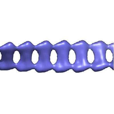 1187.gif 1187.gif | 29.2 KB | ||
| アーカイブディレクトリ |  http://ftp.pdbj.org/pub/emdb/structures/EMD-1187ftp://ftp.pdbj.org/pub/emdb/structures/EMD-1187 http://ftp.pdbj.org/pub/emdb/structures/EMD-1187ftp://ftp.pdbj.org/pub/emdb/structures/EMD-1187 | HTTPS FTP |
-関連構造データ

-リンク
| EMDBのページ | EMDB (EBI/PDBe) / EMDataResource |
|---|---|
| 「今月の分子」の関連する項目 |

-マップ
| ファイル | ダウンロード / ファイル: emd_1187.map.gz / 形式: CCP4 / 大きさ: 618.2 KB / タイプ: IMAGE STORED AS FLOATING POINT NUMBER (4 BYTES) | ||||||||||||||||||||||||||||||||||||||||||||||||||||||||||||||||||||
|---|---|---|---|---|---|---|---|---|---|---|---|---|---|---|---|---|---|---|---|---|---|---|---|---|---|---|---|---|---|---|---|---|---|---|---|---|---|---|---|---|---|---|---|---|---|---|---|---|---|---|---|---|---|---|---|---|---|---|---|---|---|---|---|---|---|---|---|---|---|
| 注釈 | Volume (map) file of a Mouse PrP amyloid fibril (900 Angstrom helical crossover repeat). | ||||||||||||||||||||||||||||||||||||||||||||||||||||||||||||||||||||
| 投影像・断面図 | 画像のコントロール
画像は Spider により作成 これらの図は立方格子座標系で作成されたものです | ||||||||||||||||||||||||||||||||||||||||||||||||||||||||||||||||||||
| ボクセルのサイズ | X=Y=Z: 5.2 Å | ||||||||||||||||||||||||||||||||||||||||||||||||||||||||||||||||||||
| 密度 |
| ||||||||||||||||||||||||||||||||||||||||||||||||||||||||||||||||||||
| 対称性 | 空間群: 1 | ||||||||||||||||||||||||||||||||||||||||||||||||||||||||||||||||||||
| 詳細 | EMDB XML:
CCP4マップ ヘッダ情報:
| ||||||||||||||||||||||||||||||||||||||||||||||||||||||||||||||||||||
 Z (Sec.)
 Y (Row.)
 X (Col.)


-添付データ
- 試料の構成要素
-全体 : Mouse Prion Protein PrP - residue 91-231
| 全体 | 名称: Mouse Prion Protein PrP - residue 91-231 |
|---|---|
| 要素 |
|
-超分子 #1000: Mouse Prion Protein PrP - residue 91-231
| 超分子 | 名称: Mouse Prion Protein PrP - residue 91-231 / タイプ: sample / ID: 1000 / 集合状態: Amyloid fibril / Number unique components: 1 |
|---|
-分子 #1: Mouse Prion Protein
| 分子 | 名称: Mouse Prion Protein / タイプ: protein_or_peptide / ID: 1 / Name.synonym: Mo PrP / 詳細: Recombinant protein expressed in E.coli / 集合状態: multimeric amyloid fibril / 組換発現: Yes |
|---|---|
| 由来(天然) | 生物種: |
| 分子量 | 実験値: 171.95 KDa / 理論値: 171.95 KDa |
| 組換発現 | 生物種:  |
-実験情報
-構造解析
| 手法 | ネガティブ染色法 |
|---|---|
 解析 解析 | 単粒子再構成法 |
| 試料の集合状態 | particle |
-試料調製
| 濃度 | 1.2 mg/mL |
|---|---|
| 緩衝液 | pH: 3 / 詳細: 10mM Tris, 10mM Sodium Acetate |
| 染色 | タイプ: NEGATIVE 詳細: 3ul sample applied to grid for 2-3 minutes before blotting. 3ul 2% uranyl acetate added and blotted after 3 minutes. |
| グリッド | 詳細: 300 mesh copper grid |
| 凍結 | 凍結剤: NONE |
- 電子顕微鏡法
| 顕微鏡 | FEI TECNAI 10 |
|---|---|
| 温度 | 平均: 293 K |
| アライメント法 | Legacy - 非点収差: objective lens astigmatism was corrected at 150,000 magnification |
| 詳細 | Low Dose. FEI TECNAI 10 microscope. Gatan single tilt negative stain holder. |
| 撮影 | カテゴリ: FILM / フィルム・検出器のモデル: KODAK SO-163 FILM / デジタル化 - スキャナー: ZEISS SCAI / デジタル化 - サンプリング間隔: 7 µm / 実像数: 477 / 平均電子線量: 10 e/Å2 / Od range: 2.5 / ビット/ピクセル: 8 |
| 電子線 | 加速電圧: 100 kV / 電子線源: TUNGSTEN HAIRPIN |
| 電子光学系 | 照射モード: FLOOD BEAM / 撮影モード: BRIGHT FIELD / Cs: 2.2 mm / 最大 デフォーカス(公称値): 1.0 µm / 最小 デフォーカス(公称値): 0.3 µm / 倍率(公称値): 27000 |
| 試料ステージ | 試料ホルダー: Eucentric / 試料ホルダーモデル: OTHER |
-画像解析
| 詳細 | Particles were selected manually. |
|---|---|
| 最終 再構成 | アルゴリズム: OTHER / 解像度のタイプ: BY AUTHOR / 解像度: 28.0 Å / 解像度の算出法: FSC 0.5 CUT-OFF / ソフトウェア - 名称: SPIDER 詳細: Crossover repeats of the disordered helical fibrils were treated as single particles 使用した粒子像数: 477 |
| 最終 2次元分類 | クラス数: 1 |
