 Movie
Movie Controller

[English] 日本語
 Yorodumi
- EMDB-1186: Elongated oligomers assemble into mammalian PrP amyloid fibrils. -
+ Open data

- Basic information
| Entry | Database: EMDB / ID: EMD-1186 | |||||||||
|---|---|---|---|---|---|---|---|---|---|---|
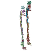
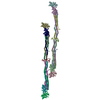
| Title | Elongated oligomers assemble into mammalian PrP amyloid fibrils. | |||||||||
 Map data Map data | Volume (map) file of a Mouse PrP amyloid fibril (1100 Angstrom helical crossover repeat). | |||||||||
 Sample Sample |
| |||||||||
| Biological species |  | |||||||||
| Method | single particle reconstruction / cryo EM / negative staining / Resolution: 26.0 Å | |||||||||
 Authors Authors | Tattum MH / Cohen-Krausz S / Khalili-shirazi A / Jackson GS / Orlova EV / Collinge J / Clarke AR / Saibil HR | |||||||||
 Citation Citation | Journal: J Mol Biol / Year: 2006 Title: Elongated oligomers assemble into mammalian PrP amyloid fibrils. Authors: M Howard Tattum / Sara Cohen-Krausz / Kanjana Thumanu / Christopher W Wharton / Azadeh Khalili-Shirazi / Graham S Jackson / Elena V Orlova / John Collinge / Anthony R Clarke / Helen R Saibil /  Abstract: In prion diseases, the mammalian prion protein PrP is converted from a monomeric, mainly alpha-helical state into beta-rich amyloid fibrils. To examine the structure of the misfolded state, amyloid ...In prion diseases, the mammalian prion protein PrP is converted from a monomeric, mainly alpha-helical state into beta-rich amyloid fibrils. To examine the structure of the misfolded state, amyloid fibrils were grown from a beta form of recombinant mouse PrP (residues 91-231). The beta-PrP precursors assembled slowly into amyloid fibrils with an overall helical twist. The fibrils exhibit immunological reactivity similar to that of ex vivo PrP Sc. Using electron microscopy and image processing, we obtained three-dimensional density maps of two forms of PrP fibrils with slightly different twists. They reveal two intertwined protofilaments with a subunit repeat of approximately 60 A. The repeating unit along each protofilament can be accounted for by elongated oligomers of PrP, suggesting a hierarchical assembly mechanism for the fibrils. The structure reveals flexible crossbridges between the two protofilaments, and subunit contacts along the protofilaments that are likely to reflect specific features of the PrP sequence, in addition to the generic, cross-beta amyloid fold. | |||||||||
| History |
|
- Structure visualization
| Movie |
 Movie viewer Movie viewer |
|---|---|
| Structure viewer | EM map: SurfViewMolmilJmol/JSmol |
| Supplemental images |
 UCSF Chimera
- Downloads & links
-EMDB archive
| Map data | emd_1186.map.gz | 584.6 KB | EMDB map data format | |
|---|---|---|---|---|
| Header (meta data) | emd-1186-v30.xmlemd-1186.xml | 10.2 KB 10.2 KB | Display Display | EMDB header |
| Images |  1186.gif 1186.gif | 24.9 KB | ||
| Archive directory |  http://ftp.pdbj.org/pub/emdb/structures/EMD-1186ftp://ftp.pdbj.org/pub/emdb/structures/EMD-1186 http://ftp.pdbj.org/pub/emdb/structures/EMD-1186ftp://ftp.pdbj.org/pub/emdb/structures/EMD-1186 | HTTPS FTP |
-Related structure data


-Links
| EMDB pages | EMDB (EBI/PDBe) / EMDataResource |
|---|---|
| Related items in Molecule of the Month |

-Map
| File | Download / File: emd_1186.map.gz / Format: CCP4 / Size: 618.2 KB / Type: IMAGE STORED AS FLOATING POINT NUMBER (4 BYTES) | ||||||||||||||||||||||||||||||||||||||||||||||||||||||||||||||||||||
|---|---|---|---|---|---|---|---|---|---|---|---|---|---|---|---|---|---|---|---|---|---|---|---|---|---|---|---|---|---|---|---|---|---|---|---|---|---|---|---|---|---|---|---|---|---|---|---|---|---|---|---|---|---|---|---|---|---|---|---|---|---|---|---|---|---|---|---|---|---|
| Annotation | Volume (map) file of a Mouse PrP amyloid fibril (1100 Angstrom helical crossover repeat). | ||||||||||||||||||||||||||||||||||||||||||||||||||||||||||||||||||||
| Projections & slices | Image control
Images are generated by Spider. generated in cubic-lattice coordinate | ||||||||||||||||||||||||||||||||||||||||||||||||||||||||||||||||||||
| Voxel size | X=Y=Z: 5.2 Å | ||||||||||||||||||||||||||||||||||||||||||||||||||||||||||||||||||||
| Density |
| ||||||||||||||||||||||||||||||||||||||||||||||||||||||||||||||||||||
| Symmetry | Space group: 1 | ||||||||||||||||||||||||||||||||||||||||||||||||||||||||||||||||||||
| Details | EMDB XML:
CCP4 map header:
| ||||||||||||||||||||||||||||||||||||||||||||||||||||||||||||||||||||
 Z (Sec.)
 Y (Row.)
 X (Col.)


-Supplemental data
- Sample components
-Entire : Mouse Prion Protein PrP - residue 91-231
| Entire | Name: Mouse Prion Protein PrP - residue 91-231 |
|---|---|
| Components |
|
-Supramolecule #1000: Mouse Prion Protein PrP - residue 91-231
| Supramolecule | Name: Mouse Prion Protein PrP - residue 91-231 / type: sample / ID: 1000 / Oligomeric state: Amyloid fibril / Number unique components: 1 |
|---|
-Macromolecule #1: Mouse Prion Protein
| Macromolecule | Name: Mouse Prion Protein / type: protein_or_peptide / ID: 1 / Name.synonym: Mo PrP / Details: Recombinant protein expressed in E.coli / Oligomeric state: multimeric amyloid fibril / Recombinant expression: Yes |
|---|---|
| Source (natural) | Organism: |
| Molecular weight | Experimental: 171.95 KDa / Theoretical: 171.95 KDa |
| Recombinant expression | Organism:  |
-Experimental details
-Structure determination
| Method | negative staining, cryo EM |
|---|---|
 Processing Processing | single particle reconstruction |
| Aggregation state | particle |
-Sample preparation
| Concentration | 1.2 mg/mL |
|---|---|
| Buffer | pH: 3 / Details: 10mM Tris, 10mM Sodium Acetate |
| Staining | Type: NEGATIVE Details: 3ul sample applied to grid for 2-3 minutes before blotting. 3ul 2% uranyl acetate added and blotted after 3 minutes. |
| Grid | Details: 300 mesh copper grid |
| Vitrification | Cryogen name: ETHANE |
- Electron microscopy
| Microscope | FEI TECNAI 10 |
|---|---|
| Temperature | Average: 298 K |
| Alignment procedure | Legacy - Astigmatism: objective lens astigmatism was corrected at |
| Details | Low dose. FEI TECNAI 10 MICROSCOPE. Gatan single tilt negative stain holder. |
| Image recording | Category: FILM / Film or detector model: KODAK SO-163 FILM / Digitization - Scanner: ZEISS SCAI / Digitization - Sampling interval: 7 µm / Number real images: 621 / Average electron dose: 10 e/Å2 / Od range: 2.5 / Bits/pixel: 8 |
| Electron beam | Acceleration voltage: 100 kV / Electron source: TUNGSTEN HAIRPIN |
| Electron optics | Illumination mode: FLOOD BEAM / Imaging mode: BRIGHT FIELD / Cs: 2.2 mm / Nominal defocus max: 1.0 µm / Nominal defocus min: 0.3 µm / Nominal magnification: 27000 |
| Sample stage | Specimen holder: eucentric / Specimen holder model: OTHER |
-Image processing
| Details | Particles were selected manually. |
|---|---|
| Final reconstruction | Algorithm: OTHER / Resolution.type: BY AUTHOR / Resolution: 26.0 Å / Resolution method: FSC 0.5 CUT-OFF / Software - Name: SPIDER Details: Crossover repeats of the disordered helical fibrils were treated as single particles Number images used: 628 |
| Final two d classification | Number classes: 1 |
