ジャーナル: Nat Commun / 年: 2020 タイトル: SARS-CoV-2 structure and replication characterized by in situ cryo-electron tomography. 著者: Steffen Klein / Mirko Cortese / Sophie L Winter / Moritz Wachsmuth-Melm / Christopher J Neufeldt / Berati Cerikan / Megan L Stanifer / Steeve Boulant / Ralf Bartenschlager / Petr Chlanda / 要旨: Severe acute respiratory syndrome coronavirus 2 (SARS-CoV-2), the causative agent of the COVID19 pandemic, is a highly pathogenic β-coronavirus. As other coronaviruses, SARS-CoV-2 is enveloped, ...Severe acute respiratory syndrome coronavirus 2 (SARS-CoV-2), the causative agent of the COVID19 pandemic, is a highly pathogenic β-coronavirus. As other coronaviruses, SARS-CoV-2 is enveloped, replicates in the cytoplasm and assembles at intracellular membranes. Here, we structurally characterize the viral replication compartment and report critical insights into the budding mechanism of the virus, and the structure of extracellular virions close to their native state by in situ cryo-electron tomography and subtomogram averaging. We directly visualize RNA filaments inside the double membrane vesicles, compartments associated with viral replication. The RNA filaments show a diameter consistent with double-stranded RNA and frequent branching likely representing RNA secondary structures. We report that assembled S trimers in lumenal cisternae do not alone induce membrane bending but laterally reorganize on the envelope during virion assembly. The viral ribonucleoprotein complexes (vRNPs) are accumulated at the curved membrane characteristic for budding sites suggesting that vRNP recruitment is enhanced by membrane curvature. Subtomogram averaging shows that vRNPs are distinct cylindrical assemblies. We propose that the genome is packaged around multiple separate vRNP complexes, thereby allowing incorporation of the unusually large coronavirus genome into the virion while maintaining high steric flexibility between the vRNPs.
生物種: Severe acute respiratory syndrome coronavirus 2 (ウイルス)
-
実験情報
-
構造解析
手法
クライオ電子顕微鏡法
解析
電子線トモグラフィー法
試料の集合状態
cell
-
試料調製
緩衝液
pH: 7
凍結
凍結剤: ETHANE / チャンバー内湿度: 70 % / チャンバー内温度: 24 K / 装置: LEICA EM GP
切片作成
集束イオンビーム - 装置: OTHER / 集束イオンビーム - イオン: OTHER / 集束イオンビーム - 電圧: 30 kV / 集束イオンビーム - 電流: 0.03 nA / 集束イオンビーム - 時間: 300 sec. / 集束イオンビーム - 温度: 93 K / 集束イオンビーム - Initial thickness: 4000 nm / 集束イオンビーム - 最終 厚さ: 150 nm 集束イオンビーム - 詳細: The value given for _emd_sectioning_focused_ion_beam.instrument is Aquilos. This is not in a list of allowed values set(['DB235', 'OTHER']) so OTHER is written into the XML file.
-
電子顕微鏡法
顕微鏡
FEI TITAN KRIOS
撮影
フィルム・検出器のモデル: GATAN K3 BIOQUANTUM (6k x 4k) 平均電子線量: 3.0 e/Å2
 ムービー
ムービー コントローラー


 データを開く
 基本情報
基本情報 マップデータ
 試料
試料
 Severe acute respiratory syndrome coronavirus 2 (ウイルス)
 データ登録者
 ドイツ, 1件
  引用
引用 構造の表示
構造の表示 ムービービューア
 ダウンロードとリンク
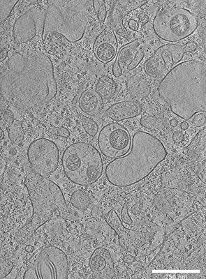 emd_11865.png
 http://ftp.pdbj.org/pub/emdb/structures/EMD-11865
 試料の構成要素
 解析
解析 電子顕微鏡法
 FIELD EMISSION GUN
