 Movie
Movie Controller

[English] 日本語
 Yorodumi
- EMDB-11863: Cryo-ET of SARS-CoV-2 infected VeroE6 cells showing budding virions. -
+ Open data

- Basic information
| Entry | Database: EMDB / ID: EMD-11863 | |||||||||
|---|---|---|---|---|---|---|---|---|---|---|
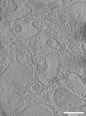
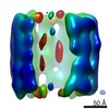
| Title | Cryo-ET of SARS-CoV-2 infected VeroE6 cells showing budding virions. | |||||||||
 Map data Map data | ||||||||||
 Sample Sample |
| |||||||||
| Biological species |   Severe acute respiratory syndrome coronavirus 2 Severe acute respiratory syndrome coronavirus 2 | |||||||||
| Method | electron tomography / cryo EM | |||||||||
 Authors Authors | Klein S / Cortese M / Winter SL / Wachsmuth-Melm M / Neufeldt CJ / Cerikan B / Stanifer ML / Boulant S / Bartenschlager R / Chlanda P | |||||||||
| Funding support |  Germany, 1 items Germany, 1 items
| |||||||||
 Citation Citation | Journal: Nat Commun / Year: 2020 Title: SARS-CoV-2 structure and replication characterized by in situ cryo-electron tomography. Authors: Steffen Klein / Mirko Cortese / Sophie L Winter / Moritz Wachsmuth-Melm / Christopher J Neufeldt / Berati Cerikan / Megan L Stanifer / Steeve Boulant / Ralf Bartenschlager / Petr Chlanda / Abstract: Severe acute respiratory syndrome coronavirus 2 (SARS-CoV-2), the causative agent of the COVID19 pandemic, is a highly pathogenic β-coronavirus. As other coronaviruses, SARS-CoV-2 is enveloped, ...Severe acute respiratory syndrome coronavirus 2 (SARS-CoV-2), the causative agent of the COVID19 pandemic, is a highly pathogenic β-coronavirus. As other coronaviruses, SARS-CoV-2 is enveloped, replicates in the cytoplasm and assembles at intracellular membranes. Here, we structurally characterize the viral replication compartment and report critical insights into the budding mechanism of the virus, and the structure of extracellular virions close to their native state by in situ cryo-electron tomography and subtomogram averaging. We directly visualize RNA filaments inside the double membrane vesicles, compartments associated with viral replication. The RNA filaments show a diameter consistent with double-stranded RNA and frequent branching likely representing RNA secondary structures. We report that assembled S trimers in lumenal cisternae do not alone induce membrane bending but laterally reorganize on the envelope during virion assembly. The viral ribonucleoprotein complexes (vRNPs) are accumulated at the curved membrane characteristic for budding sites suggesting that vRNP recruitment is enhanced by membrane curvature. Subtomogram averaging shows that vRNPs are distinct cylindrical assemblies. We propose that the genome is packaged around multiple separate vRNP complexes, thereby allowing incorporation of the unusually large coronavirus genome into the virion while maintaining high steric flexibility between the vRNPs. #1: Journal: Biorxiv / Year: 2020Title: SARS-CoV-2 structure and replication characterized by in situ cryo-electron tomography Authors: Klein S / Cortese M / Winter SL / Wachsmuth-Melm M / Neufeldt CJ / Cerikan B / Stanifer ML / Boulant S / Bartenschlager R / Chlanda P | |||||||||
| History |
|
- Structure visualization
| Movie |
 Movie viewer Movie viewer |
|---|---|
| Supplemental images |
- Downloads & links
-EMDB archive
| Map data | emd_11863.map.gz | 451.6 MB | EMDB map data format | |
|---|---|---|---|---|
| Header (meta data) | emd-11863-v30.xmlemd-11863.xml | 10.2 KB 10.2 KB | Display Display | EMDB header |
| Images |  emd_11863.png emd_11863.png | 100.8 KB | ||
| Archive directory |  http://ftp.pdbj.org/pub/emdb/structures/EMD-11863ftp://ftp.pdbj.org/pub/emdb/structures/EMD-11863 http://ftp.pdbj.org/pub/emdb/structures/EMD-11863ftp://ftp.pdbj.org/pub/emdb/structures/EMD-11863 | HTTPS FTP |
-Related structure data
-Links
| EMDB pages | EMDB (EBI/PDBe) / EMDataResource |
|---|
-Map
| File | Download / File: emd_11863.map.gz / Format: CCP4 / Size: 594.4 MB / Type: IMAGE STORED AS SIGNED BYTE | ||||||||||||||||||||||||||||||||||||||||||||||||||||||||||||||||||||
|---|---|---|---|---|---|---|---|---|---|---|---|---|---|---|---|---|---|---|---|---|---|---|---|---|---|---|---|---|---|---|---|---|---|---|---|---|---|---|---|---|---|---|---|---|---|---|---|---|---|---|---|---|---|---|---|---|---|---|---|---|---|---|---|---|---|---|---|---|---|
| Voxel size | X=Y=Z: 8.013 Å | ||||||||||||||||||||||||||||||||||||||||||||||||||||||||||||||||||||
| Density |
| ||||||||||||||||||||||||||||||||||||||||||||||||||||||||||||||||||||
| Symmetry | Space group: 1 | ||||||||||||||||||||||||||||||||||||||||||||||||||||||||||||||||||||
| Details | EMDB XML:
CCP4 map header:
| ||||||||||||||||||||||||||||||||||||||||||||||||||||||||||||||||||||
-Supplemental data
- Sample components
-Entire : VeroE6 cells infected with SARS-CoV-2
| Entire | Name: VeroE6 cells infected with SARS-CoV-2 |
|---|---|
| Components |
|
-Supramolecule #1: VeroE6 cells infected with SARS-CoV-2
| Supramolecule | Name: VeroE6 cells infected with SARS-CoV-2 / type: cell / ID: 1 / Parent: 0 |
|---|---|
| Source (natural) | Organism: Severe acute respiratory syndrome coronavirus 2 |
-Experimental details
-Structure determination
| Method | cryo EM |
|---|---|
 Processing Processing | electron tomography |
| Aggregation state | cell |
-Sample preparation
| Buffer | pH: 7 |
|---|---|
| Grid | Model: Quantifoil R2/2 / Material: GOLD / Mesh: 200 |
| Vitrification | Cryogen name: ETHANE / Chamber humidity: 70 % / Chamber temperature: 24 K / Instrument: LEICA EM GP |
| Sectioning | Focused ion beam - Instrument: OTHER / Focused ion beam - Ion: OTHER / Focused ion beam - Voltage: 30 kV / Focused ion beam - Current: 0.03 nA / Focused ion beam - Duration: 300 sec. / Focused ion beam - Temperature: 93 K / Focused ion beam - Initial thickness: 4000 nm / Focused ion beam - Final thickness: 150 nm Focused ion beam - Details: The value given for _emd_sectioning_focused_ion_beam.instrument is Aquilos. This is not in a list of allowed values set(['DB235', 'OTHER']) so OTHER is written into the XML file. |
- Electron microscopy
| Microscope | FEI TITAN KRIOS |
|---|---|
| Image recording | Film or detector model: GATAN K3 BIOQUANTUM (6k x 4k) / Average electron dose: 3.0 e/Å2 |
| Electron beam | Acceleration voltage: 300 kV / Electron source:  FIELD EMISSION GUN FIELD EMISSION GUN |
| Electron optics | C2 aperture diameter: 70.0 µm / Illumination mode: FLOOD BEAM / Imaging mode: BRIGHT FIELD |
| Experimental equipment |  Model: Titan Krios / Image courtesy: FEI Company |
-Image processing
| Final reconstruction | Number images used: 41 |
|---|
