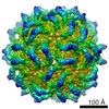
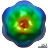
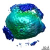
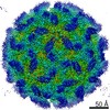
[日本語] English
- EMDB-1178: Structure of the dodecahedral penton particle from human adenovir... -

+
データを開く


IDまたはキーワード:

読み込み中...

-
基本情報

登録情報
データベース: EMDB / ID: EMD-1178
タイトルStructure of the dodecahedral penton particle from human adenovirus type 3.
マップデータThe pixel size has been calibrated using the X-ray structure of adenovirus 2 penton base. Accession number 1X9P
試料
  • 試料: adenovirus 3 penton base dodecahedron
  • タンパク質・ペプチド: Ad3 penton base
機能・相同性
機能・相同性情報


T=25 icosahedral viral capsid / adhesion receptor-mediated virion attachment to host cell / viral capsid / clathrin-dependent endocytosis of virus by host cell / cell adhesion / symbiont entry into host cell / host cell nucleus / virion attachment to host cell / structural molecule activity
類似検索 - 分子機能
Adenovirus penton base protein / Adenovirus penton base protein / Adenoviral fibre protein, knob / Adenoviral fibre protein (knob domain) / Adenoviral fibre protein, repeat/shaft region / Adenoviral fibre protein (repeat/shaft region) / Adenovirus fibre protein / Attachment protein shaft domain superfamily / Adenovirus pIV-like, attachment domain
類似検索 - ドメイン・相同性
Fiber protein / Penton protein / L5 fiber
類似検索 - 構成要素
生物種Human adenovirus 3 (ヒトアデノウイルス)
手法単粒子再構成法 / クライオ電子顕微鏡法 / ネガティブ染色法 / 解像度: 9.3 Å
データ登録者Fuschiotti P / Schoehn G / Fender P / Fabry CMS / Hewat EA / Chroboczek J
引用ジャーナル: J Mol Biol / : 2006
タイトル: Structure of the dodecahedral penton particle from human adenovirus type 3.
著者: P Fuschiotti / G Schoehn / P Fender / C M S Fabry / E A Hewat / J Chroboczek / R W H Ruigrok / J F Conway /
要旨: The sub-viral dodecahedral particle of human adenovirus type 3, composed of the viral penton base and fiber proteins, shares an important characteristic of the entire virus: it can attach to cells ...The sub-viral dodecahedral particle of human adenovirus type 3, composed of the viral penton base and fiber proteins, shares an important characteristic of the entire virus: it can attach to cells and penetrate them. Structure determination of the fiberless dodecahedron by cryo-electron microscopy to 9 Angstroms resolution reveals tightly bound pentamer subunits, with only minimal interfaces between penton bases stabilizing the fragile dodecahedron. The internal cavity of the dodecahedron is approximately 80 Angstroms in diameter, and the interior surface is accessible to solvent through perforations of approximately 20 Angstroms diameter between the pentamer towers. We observe weak density beneath pentamers that we attribute to a penton base peptide including residues 38-48. The intact amino-terminal domain appears to interfere with pentamer-pentamer interactions and its absence by mutation or proteolysis is essential for dodecamer assembly. Differences between the 9 Angstroms dodecahedron structure and the adenovirus serotype 2 (Ad2) crystallographic model correlate closely with differences in sequence. The 3D structure of the dodecahedron including fibers at 16 Angstroms resolution reveals extra density on the top of the penton base that can be attributed to the fiber N terminus. The fiber itself exhibits striations that correlate with features of the atomic structure of the partial Ad2 fiber and that represent a repeat motif present in the amino acid sequence. These new observations offer important insights into particle assembly and stability, as well as the practicality of using the dodecahedron in targeted drug delivery. The structural work provides a sound basis for manipulating the properties of this particle and thereby enhancing its value for such therapeutic use.
履歴
登録2005年11月4日-
ヘッダ(付随情報) 公開2005年11月4日-
マップ公開2005年11月7日-
更新2012年10月24日-
現状2012年10月24日処理サイト: PDBe / 状態: 公開

-
構造の表示

ムービー
  • 表面図(断面を密度値に従い着色)
  • 表面レベル: 110
  • UCSF Chimeraによる作画
  • ダウンロード
  • 表面図(半径に従い着色)
  • 表面レベル: 110
  • UCSF Chimeraによる作画
  • ダウンロード
  • あてはめたモデルとの重ね合わせ
  • 原子モデル: PDB-2c9g
  • 表面レベル: 110
  • UCSF Chimeraによる作画
  • ダウンロード
  • 単純化した表面モデル + あてはめた原子モデル
  • 原子モデルPDB-2c9g
  • Jmolによる作画
  • ダウンロード
ムービービューア
構造ビューアEMマップ:
SurfViewMolmilJmol/JSmol
添付画像

ダウンロードとリンク

-
マップ

ファイルダウンロード / ファイル: emd_1178.map.gz / 形式: CCP4 / 大きさ: 40.5 MB / タイプ: IMAGE STORED AS SIGNED INTEGER (2 BYTES)
注釈The pixel size has been calibrated using the X-ray structure of adenovirus 2 penton base. Accession number 1X9P
ボクセルのサイズX=Y=Z: 1.37 Å
密度
表面レベル登録者による: 91.900000000000006 / ムービー #1: 110
最小 - 最大-335.0 - 509.0
平均 (標準偏差)3.05461431 (±52.415409089999997)
対称性空間群: 1
詳細

EMDB XML:

マップ形状
Axis orderXYZ
Origin-139-139-139
サイズ279279279
Spacing279279279
セルA=B=C: 382.23 Å
α=β=γ: 90.0 °

CCP4マップ ヘッダ情報:

modeImage stored as Integer*27
Å/pix. X/Y/Z1.371.371.37
M x/y/z279279279
origin x/y/z0.0000.0000.000
length x/y/z382.230382.230382.230
α/β/γ90.00090.00090.000
start NX/NY/NZ-77-770
NX/NY/NZ15515578
MAP C/R/S123
start NC/NR/NS-139-139-139
NC/NR/NS279279279
D min/max/mean-335.000509.0003.055

-
添付データ

-
試料の構成要素

-
全体 : adenovirus 3 penton base dodecahedron

全体名称: adenovirus 3 penton base dodecahedron
要素
  • 試料: adenovirus 3 penton base dodecahedron
  • タンパク質・ペプチド: Ad3 penton base

-
超分子 #1000: adenovirus 3 penton base dodecahedron

超分子名称: adenovirus 3 penton base dodecahedron / タイプ: sample / ID: 1000
詳細: The penton base was expressed in baculovirus abd the penton base self-assemble into dodecahedrons
集合状態: 12 pentamers / Number unique components: 1
分子量実験値: 3.5 MDa

-
分子 #1: Ad3 penton base

分子名称: Ad3 penton base / タイプ: protein_or_peptide / ID: 1 / コピー数: 60 / 集合状態: dodecamer of pentamer / 組換発現: Yes
由来(天然)生物種: Human adenovirus 3 (ヒトアデノウイルス) / 別称: human adenovirus 3
分子量実験値: 60 KDa / 理論値: 60 KDa
組換発現生物種: unidentified baculovirus (ウイルス) / 組換プラスミド: pAcUW31

-
実験情報

-
構造解析

手法ネガティブ染色法, クライオ電子顕微鏡法
解析単粒子再構成法
試料の集合状態particle

-
試料調製

濃度1 mg/mL
緩衝液pH: 6.6 / 詳細: 25 mM phosphate buffer at pH 6.6
染色タイプ: NEGATIVE
詳細: Quantifoil R2 1 grids (Quantifoil Micro Tools GmbH, Germany) were loaded with 4 ul of sample at 1 mg ml, blotted and rapidly frozen in liquid ethane within a liquid nitrogen bath using a Zeiss cryoplunger
グリッド詳細: Quantifoil R2/1 grids
凍結凍結剤: ETHANE / チャンバー内温度: 100 K / 装置: OTHER / 詳細: Vitrification instrument: Zeiss cryoplunger

-
電子顕微鏡法

顕微鏡JEOL 2010F
温度平均: 100 K
アライメント法Legacy - 非点収差: objective lens astigmatism was corrected at 100,000
撮影カテゴリ: FILM / フィルム・検出器のモデル: KODAK SO-163 FILM / デジタル化 - スキャナー: ZEISS SCAI / デジタル化 - サンプリング間隔: 7 µm / 実像数: 14
電子線加速電圧: 200 kV / 電子線源: FIELD EMISSION GUN
電子光学系倍率(補正後): 51100 / 照射モード: OTHER / 撮影モード: BRIGHT FIELD / Cs: 1.4 mm / 最大 デフォーカス(公称値): 2.5 µm / 最小 デフォーカス(公称値): 1.0 µm / 倍率(公称値): 50000
試料ステージ試料ホルダー: Eucentric / 試料ホルダーモデル: OTHER

-
画像解析

詳細The particles were selected using X3d
CTF補正詳細: each negative or each focal pair
最終 再構成想定した対称性 - 点群: I (正20面体型対称) / アルゴリズム: OTHER / 解像度のタイプ: BY AUTHOR / 解像度: 9.3 Å / 解像度の算出法: FSC 0.33 CUT-OFF / ソフトウェア - 名称: PFT EM3DR PFT2 EM3DR2 / 使用した粒子像数: 1849

+
万見について

-
お知らせ

-
2022年2月9日: EMDBエントリの付随情報ファイルのフォーマットが新しくなりました

EMDBエントリの付随情報ファイルのフォーマットが新しくなりました

  • EMDBのヘッダファイルのバージョン3が、公式のフォーマットとなりました。
  • これまでは公式だったバージョン1.9は、アーカイブから削除されます。

関連情報:EMDBヘッダ

外部リンク:wwPDBはEMDBデータモデルのバージョン3へ移行します

-
2020年8月12日: 新型コロナ情報

新型コロナ情報

URL: https://pdbjlvh1.pdbj.org/emnavi/covid19.php

新ページ: EM Navigatorに新型コロナウイルスの特設ページを開設しました。

関連情報:Covid-19情報 / 2020年3月5日: 新型コロナウイルスの構造データ

+
2020年3月5日: 新型コロナウイルスの構造データ

新型コロナウイルスの構造データ

関連情報:万見生物種 / 2020年8月12日: 新型コロナ情報

外部リンク:COVID-19特集ページ - PDBj / 今月の分子2020年2月:コロナウイルスプロテーアーゼ

+
2019年1月31日: EMDBのIDの桁数の変更

EMDBのIDの桁数の変更

  • EMDBエントリに付与されているアクセスコード(EMDB-ID)は4桁の数字(例、EMD-1234)でしたが、間もなく枯渇します。これまでの4桁のID番号は4桁のまま変更されませんが、4桁の数字を使い切った後に発行されるIDは5桁以上の数字(例、EMD-12345)になります。5桁のIDは2019年の春頃から発行される見通しです。
  • EM Navigator/万見では、接頭語「EMD-」は省略されています。

関連情報:Q: 「EMD」とは何ですか? / 万見/EM NavigatorにおけるID/アクセスコードの表記

外部リンク:EMDB Accession Codes are Changing Soon! / PDBjへお問い合わせ

+
2017年7月12日: PDB大規模アップデート

PDB大規模アップデート

  • 新バージョンのPDBx/mmCIF辞書形式に基づくデータがリリースされました。
  • 今回の更新はバージョン番号が4から5になる大規模なもので、全エントリデータの書き換えが行われる「Remediation」というアップデートに該当します。
  • このバージョンアップで、電子顕微鏡の実験手法に関する多くの項目の書式が改定されました(例:em_softwareなど)。
  • EM NavigatorとYorodumiでも、この改定に基づいた表示内容になります。

外部リンク:wwPDB Remediation / OneDepデータ基準に準拠した、より強化された内容のモデル構造ファイルが、PDBアーカイブで公開されました。

-
万見 (Yorodumi)

幾万の構造データを、幾万の視点から

  • 万見(Yorodumi)は、EMDB/PDB/SASBDBなどの構造データを閲覧するためのページです。
  • EM Navigatorの詳細ページの後継、Omokage検索のフロントエンドも兼ねています。

関連情報:EMDB / PDB / SASBDB / 3つのデータバンクの比較 / 万見検索 / 2016年8月31日: 新しいEM Navigatorと万見 / 万見文献 / Jmol/JSmol / 機能・相同性情報 / 新しいEM Navigatorと万見の変更点

他の情報も見る