[English] 日本語
 Yorodumi
- EMDB-1177: Structure of Broadhaven virus by cryoelectron microscopy: correla... -
+ Open data
- Basic information
| Entry | Database: EMDB / ID: EMD-1177 | |||||||||
|---|---|---|---|---|---|---|---|---|---|---|
| Title | Structure of Broadhaven virus by cryoelectron microscopy: correlation of structural and antigenic properties of Broadhaven virus and bluetongue virus outer capsid proteins. | |||||||||
 Map data Map data | 3d reconstruction with phase flipped images | |||||||||
 Sample Sample |
| |||||||||
| Biological species |  Broadhaven virus Broadhaven virus | |||||||||
| Method | single particle reconstruction / cryo EM / negative staining / Resolution: 23.0 Å | |||||||||
 Authors Authors | Schoehn G / Moss SR / Nuttall PA / Hewat EA | |||||||||
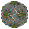
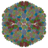
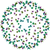
 Citation Citation |  Journal: Virology / Year: 1997 Journal: Virology / Year: 1997Title: Structure of Broadhaven virus by cryoelectron microscopy: correlation of structural and antigenic properties of Broadhaven virus and bluetongue virus outer capsid proteins. Authors: G Schoehn / S R Moss / P A Nuttall / E A Hewat /  Abstract: The three-dimensional structure of Broadhaven virus (BRDV) has been determined to 23 A resolution by cryoelectron microscopy and image processing. As predicted from sequence homology, the BRDV ...The three-dimensional structure of Broadhaven virus (BRDV) has been determined to 23 A resolution by cryoelectron microscopy and image processing. As predicted from sequence homology, the BRDV structure resembles that of bluetongue virus (BTV) with the notable exception of one of the outer shell proteins. The cores of BRDV and BTV are identical at medium resolution; they have a diameter of 710 A and the VP7 trimers are arranged on a T = 13 icosahedral lattice. The outer shell proteins, VP5 of BRDV and BTV, have roughly the same molecular weight while VP4 of BRDV is only half the molecular weight of the corresponding VP2 of BTV. This size difference allows unambiguous determination of the identity of the triskelion shape as trimers of VP4 of BRDV (VP2 of BTV). The VP4 of BRDV sits on the VP7 trimers and projects outwards 40 A, giving the capsid an overall diameter of 790 A. This contrasts with VP2 of BTV, which projects outwards 95 A to give the capsid a diameter of 900 A. The difference in accessibility of the outer shell proteins of BRDV and BTV correlates with the difference in antigenic properties of these viral proteins. The shape of the BRDV VP5 indicates that it too is a trimer, thus implying that there are 360 copies of VP5 and 180 copies of VP4 per virion. | |||||||||
| History |
|
- Structure visualization
| Movie |
 Movie viewer Movie viewer |
|---|---|
| Structure viewer | EM map:  SurfView SurfView Molmil Molmil Jmol/JSmol Jmol/JSmol |
| Supplemental images |
- Downloads & links
-EMDB archive
| Map data |  emd_1177.map.gz emd_1177.map.gz | 17.6 MB |  EMDB map data format EMDB map data format | |
|---|---|---|---|---|
| Header (meta data) |  emd-1177-v30.xml emd-1177-v30.xml emd-1177.xml emd-1177.xml | 8.5 KB 8.5 KB | Display Display |  EMDB header EMDB header |
| Images |  1177.gif 1177.gif | 66 KB | ||
| Archive directory |  http://ftp.pdbj.org/pub/emdb/structures/EMD-1177 http://ftp.pdbj.org/pub/emdb/structures/EMD-1177 ftp://ftp.pdbj.org/pub/emdb/structures/EMD-1177 ftp://ftp.pdbj.org/pub/emdb/structures/EMD-1177 | HTTPS FTP |
-Related structure data
| Similar structure data |
|---|
- Links
Links
| EMDB pages |  EMDB (EBI/PDBe) / EMDB (EBI/PDBe) /  EMDataResource EMDataResource |
|---|
- Map
Map
| File |  Download / File: emd_1177.map.gz / Format: CCP4 / Size: 18.3 MB / Type: IMAGE STORED AS FLOATING POINT NUMBER (4 BYTES) Download / File: emd_1177.map.gz / Format: CCP4 / Size: 18.3 MB / Type: IMAGE STORED AS FLOATING POINT NUMBER (4 BYTES) | ||||||||||||||||||||||||||||||||||||||||||||||||||||||||||||||||||||
|---|---|---|---|---|---|---|---|---|---|---|---|---|---|---|---|---|---|---|---|---|---|---|---|---|---|---|---|---|---|---|---|---|---|---|---|---|---|---|---|---|---|---|---|---|---|---|---|---|---|---|---|---|---|---|---|---|---|---|---|---|---|---|---|---|---|---|---|---|---|
| Annotation | 3d reconstruction with phase flipped images | ||||||||||||||||||||||||||||||||||||||||||||||||||||||||||||||||||||
| Projections & slices | Image control
Images are generated by Spider. | ||||||||||||||||||||||||||||||||||||||||||||||||||||||||||||||||||||
| Voxel size | X=Y=Z: 5.09 Å | ||||||||||||||||||||||||||||||||||||||||||||||||||||||||||||||||||||
| Density |
| ||||||||||||||||||||||||||||||||||||||||||||||||||||||||||||||||||||
| Symmetry | Space group: 1 | ||||||||||||||||||||||||||||||||||||||||||||||||||||||||||||||||||||
| Details | EMDB XML:
CCP4 map header:
| ||||||||||||||||||||||||||||||||||||||||||||||||||||||||||||||||||||
-Supplemental data
- Sample components
-Entire : Broadhaven virus
| Entire | Name:  Broadhaven virus Broadhaven virus |
|---|---|
| Components |
|
-Supramolecule #1000: Broadhaven virus
| Supramolecule | Name: Broadhaven virus / type: sample / ID: 1000 / Details: Virus grown in BHK cells and purified / Number unique components: 1 |
|---|
-Supramolecule #1: Broadhaven virus
| Supramolecule | Name: Broadhaven virus / type: virus / ID: 1 / NCBI-ID: 10893 / Sci species name: Broadhaven virus / Virus type: VIRION / Virus isolate: STRAIN / Virus enveloped: No / Virus empty: No |
|---|---|
| Host (natural) | Organism:  sea bird (butterflies/moths) / synonym: VERTEBRATES sea bird (butterflies/moths) / synonym: VERTEBRATES |
| Virus shell | Shell ID: 1 / Name: triple shell / Diameter: 800 Å / T number (triangulation number): 13 |
-Experimental details
-Structure determination
| Method | negative staining, cryo EM |
|---|---|
 Processing Processing | single particle reconstruction |
| Aggregation state | particle |
- Sample preparation
| Concentration | 0.5 mg/mL |
|---|---|
| Staining | Type: NEGATIVE Details: Cryo EM on home made holey grids covered by a thin layer of carbon |
| Grid | Details: 400 mesh grid |
| Vitrification | Cryogen name: ETHANE / Instrument: OTHER Details: Vitrification instrument: zeiss. on holey grid covered by carbon to increase the concentration |
- Electron microscopy
| Microscope | FEI/PHILIPS CM200T |
|---|---|
| Alignment procedure | Legacy - Astigmatism: objective lens astigmatism was corrected at 100,000 |
| Date | Feb 12, 1996 |
| Image recording | Category: FILM / Film or detector model: KODAK SO-163 FILM / Digitization - Scanner: ZEISS SCAI / Digitization - Sampling interval: 14 µm / Number real images: 10 / Average electron dose: 9 e/Å2 / Bits/pixel: 8 |
| Electron beam | Acceleration voltage: 200 kV / Electron source: LAB6 |
| Electron optics | Illumination mode: OTHER / Imaging mode: OTHER / Cs: 2 mm / Nominal defocus max: 3.0 µm / Nominal defocus min: 1.0 µm / Nominal magnification: 27500 |
| Sample stage | Specimen holder: Eucentric / Specimen holder model: GATAN LIQUID NITROGEN |
- Image processing
| CTF correction | Details: Each particle |
|---|---|
| Final reconstruction | Applied symmetry - Point group: I (icosahedral) / Algorithm: OTHER / Resolution.type: BY AUTHOR / Resolution: 23.0 Å / Resolution method: OTHER / Software - Name: pft / Number images used: 200 |
 Movie
Movie Controller


 UCSF Chimera


 Z (Sec.)
 Y (Row.)
 X (Col.)


