 ムービー
ムービー コントローラー


+ データを開く

- 基本情報
基本情報
| 登録情報 | データベース: EMDB / ID: EMD-1165 | |||||||||
|---|---|---|---|---|---|---|---|---|---|---|
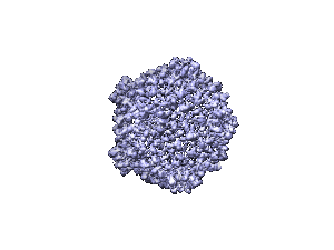
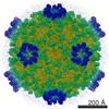
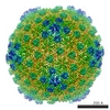
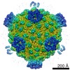
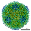
| タイトル | A 9 angstroms single particle reconstruction from CCD captured images on a 200 kV electron cryomicroscope. | |||||||||
 マップデータ マップデータ | this is a half map of CPV oriented along the 5 fold axis of symmetry. Raw Data Collected on 4k x 4k Gatan US4000 CCD Camera. Reconstruction to 9 A. | |||||||||
 試料 試料 |
| |||||||||
| 生物種 |   Bombyx mori cypovirus 1 (ウイルス) Bombyx mori cypovirus 1 (ウイルス) | |||||||||
| 手法 | 単粒子再構成法 / クライオ電子顕微鏡法 / ネガティブ染色法 / 解像度: 9.0 Å | |||||||||
 データ登録者 データ登録者 | Booth C / Jiang W / Baker ML / Zhou ZH / Ludtke SJ / Chiu W | |||||||||
 引用 引用 | ジャーナル: J Struct Biol / 年: 2004 タイトル: A 9 angstroms single particle reconstruction from CCD captured images on a 200 kV electron cryomicroscope. 著者: Christopher R Booth / Wen Jiang / Matthew L Baker / Z Hong Zhou / Steven J Ludtke / Wah Chiu /  要旨: Sub-nanometer resolution structure determination is becoming a common practice in electron cryomicroscopy of macromolecular assemblies. The data for these studies have until now been collected on ...Sub-nanometer resolution structure determination is becoming a common practice in electron cryomicroscopy of macromolecular assemblies. The data for these studies have until now been collected on photographic film. Using cytoplasmic polyhedrosis virus (CPV), a previously determined structure, as a test specimen, we show the feasibility of obtaining a 9 angstroms structure from images acquired from a 4 k x 4 k Gatan CCD on a 200 kV electron cryomicroscope. The match of the alpha-helices in the protein components of the CPV with the previous structure of the same virus validates the suitability of this type of camera as the recording media targeted for single particle reconstructions at sub-nanometer resolution. | |||||||||
| 履歴 |
|
- 構造の表示
構造の表示
| ムービー |
 ムービービューア ムービービューア |
|---|---|
| 構造ビューア | EMマップ: SurfViewMolmilJmol/JSmol |
| 添付画像 |
- ダウンロードとリンク
-EMDBアーカイブ
| マップデータ | emd_1165.map.gz | 117.9 MB | EMDBマップデータ形式 | |
|---|---|---|---|---|
| ヘッダ (付随情報) | emd-1165-v30.xmlemd-1165.xml | 9.6 KB 9.6 KB | 表示 表示 | EMDBヘッダ |
| 画像 |  1165.gif 1165.gif | 13.4 KB | ||
| アーカイブディレクトリ |  http://ftp.pdbj.org/pub/emdb/structures/EMD-1165ftp://ftp.pdbj.org/pub/emdb/structures/EMD-1165 http://ftp.pdbj.org/pub/emdb/structures/EMD-1165ftp://ftp.pdbj.org/pub/emdb/structures/EMD-1165 | HTTPS FTP |
-検証レポート
| 文書・要旨 | emd_1165_validation.pdf.gz | 248.7 KB | 表示 | EMDB検証レポート |
|---|---|---|---|---|
| 文書・詳細版 | emd_1165_full_validation.pdf.gz | 247.8 KB | 表示 | |
| XML形式データ | emd_1165_validation.xml.gz | 4.9 KB | 表示 | |
| アーカイブディレクトリ | https://ftp.pdbj.org/pub/emdb/validation_reports/EMD-1165ftp://ftp.pdbj.org/pub/emdb/validation_reports/EMD-1165 | HTTPS FTP |
-関連構造データ


-リンク
| EMDBのページ | EMDB (EBI/PDBe) / EMDataResource |
|---|
-マップ
| ファイル | ダウンロード / ファイル: emd_1165.map.gz / 形式: CCP4 / 大きさ: 231.9 MB / タイプ: IMAGE STORED AS FLOATING POINT NUMBER (4 BYTES) | ||||||||||||||||||||||||||||||||||||||||||||||||||||||||||||||||||||
|---|---|---|---|---|---|---|---|---|---|---|---|---|---|---|---|---|---|---|---|---|---|---|---|---|---|---|---|---|---|---|---|---|---|---|---|---|---|---|---|---|---|---|---|---|---|---|---|---|---|---|---|---|---|---|---|---|---|---|---|---|---|---|---|---|---|---|---|---|---|
| 注釈 | this is a half map of CPV oriented along the 5 fold axis of symmetry. Raw Data Collected on 4k x 4k Gatan US4000 CCD Camera. Reconstruction to 9 A. | ||||||||||||||||||||||||||||||||||||||||||||||||||||||||||||||||||||
| 投影像・断面図 | 画像のコントロール
画像は Spider により作成 これらの図は立方格子座標系で作成されたものです | ||||||||||||||||||||||||||||||||||||||||||||||||||||||||||||||||||||
| ボクセルのサイズ | X=Y=Z: 1.8 Å | ||||||||||||||||||||||||||||||||||||||||||||||||||||||||||||||||||||
| 密度 |
| ||||||||||||||||||||||||||||||||||||||||||||||||||||||||||||||||||||
| 対称性 | 空間群: 1 | ||||||||||||||||||||||||||||||||||||||||||||||||||||||||||||||||||||
| 詳細 | EMDB XML:
CCP4マップ ヘッダ情報:
| ||||||||||||||||||||||||||||||||||||||||||||||||||||||||||||||||||||
 Z (Sec.)
 Y (Row.)
 X (Col.)


-添付データ
- 試料の構成要素
-全体 : Cytoplasmic Polyhedrosis Virus
| 全体 | 名称: Cytoplasmic Polyhedrosis Virus |
|---|---|
| 要素 |
|
-超分子 #1000: Cytoplasmic Polyhedrosis Virus
| 超分子 | 名称: Cytoplasmic Polyhedrosis Virus / タイプ: sample / ID: 1000 / Number unique components: 1 |
|---|
-超分子 #1: Bombyx mori cypovirus 1
| 超分子 | 名称: Bombyx mori cypovirus 1 / タイプ: virus / ID: 1 / Name.synonym: CPV / NCBI-ID: 110829 / 生物種: Bombyx mori cypovirus 1 / ウイルスタイプ: VIRION / ウイルス・単離状態: SPECIES / ウイルス・エンベロープ: No / ウイルス・中空状態: No / Syn species name: CPV |
|---|---|
| 宿主 | 生物種:  |
| ウイルス殻 | Shell ID: 1 / 名称: capsid / 直径: 750 Å / T番号(三角分割数): 1 |
-実験情報
-構造解析
| 手法 | ネガティブ染色法, クライオ電子顕微鏡法 |
|---|---|
 解析 解析 | 単粒子再構成法 |
| 試料の集合状態 | particle |
-試料調製
| 濃度 | 1 mg/mL |
|---|---|
| 緩衝液 | pH: 7.4 / 詳細: Phosphate Buffered Saline ph 7.4 |
| 染色 | タイプ: NEGATIVE / 詳細: flash frozen in liquid ethane |
| グリッド | 詳細: irradiated quantafoil R2-1 holey carbon grids |
| 凍結 | 凍結剤: ETHANE / チャンバー内湿度: 45 % / チャンバー内温度: 80 K / 装置: HOMEMADE PLUNGER 詳細: Vitrification instrument: manual plunger. homemade apparatus 手法: blot on 1 side and plunge |
- 電子顕微鏡法
| 顕微鏡 | JEOL 2010F |
|---|---|
| 温度 | 最低: 90 K / 最高: 90 K / 平均: 90 K |
| 日付 | 2003年3月1日 |
| 撮影 | カテゴリ: CCD フィルム・検出器のモデル: GATAN ULTRASCAN 4000 (4k x 4k) 実像数: 856 / 平均電子線量: 15 e/Å2 / ビット/ピクセル: 12 |
| 電子線 | 加速電圧: 200 kV / 電子線源:  FIELD EMISSION GUN FIELD EMISSION GUN |
| 電子光学系 | 倍率(補正後): 83062 / 照射モード: FLOOD BEAM / 撮影モード: BRIGHT FIELD / Cs: 1 mm / 最大 デフォーカス(公称値): 4.0 µm / 最小 デフォーカス(公称値): 0.8 µm / 倍率(公称値): 60000 |
| 試料ステージ | 試料ホルダー: Side entry liquid nitrogen-cooled cryo specimen holder 試料ホルダーモデル: GATAN LIQUID NITROGEN |
-画像解析
| 詳細 | particles selected using Ethan and manually verified. |
|---|---|
| CTF補正 | 詳細: SAVR per frame |
| 最終 再構成 | 想定した対称性 - 点群: I (正20面体型対称) / アルゴリズム: OTHER / 解像度のタイプ: BY AUTHOR / 解像度: 9.0 Å / 解像度の算出法: FSC 0.5 CUT-OFF / ソフトウェア - 名称: SAVR / 使用した粒子像数: 6465 |
-原子モデル構築 1
| ソフトウェア | 名称: Foldhunter |
|---|---|
| 詳細 | Protocol: Rigid Body. Reovirus lambda1 protein was fitted to CSPA. Helixhunter was used to identify alpha helices. |
| 精密化 | プロトコル: RIGID BODY FIT / 当てはまり具合の基準: Cross correlation |
