Movie
Movie Controller

[English] 日本語
 Yorodumi
- EMDB-1141: The structure of p53 tumour suppressor protein reveals the basis ... -
+ Open data

- Basic information
| Entry | Database: EMDB / ID: EMD-1141 | |||||||||
|---|---|---|---|---|---|---|---|---|---|---|
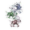
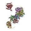
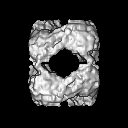
| Title | The structure of p53 tumour suppressor protein reveals the basis for its functional plasticity. | |||||||||
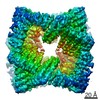
 Map data Map data | The EM map of the p53 tumour suppressor | |||||||||
 Sample Sample |
| |||||||||
| Function / homology | p53 tumour suppressor family / nucleus Function and homology information Function and homology information | |||||||||
| Biological species |  | |||||||||
| Method | single particle reconstruction / cryo EM / Resolution: 13.7 Å | |||||||||
 Authors Authors | Okorokov AL / Sherman MB / Milner J / Orlova EV | |||||||||
 Citation Citation | Journal: EMBO J / Year: 2006 Title: The structure of p53 tumour suppressor protein reveals the basis for its functional plasticity. Authors: Andrei L Okorokov / Michael B Sherman / Celia Plisson / Vera Grinkevich / Kristmundur Sigmundsson / Galina Selivanova / Jo Milner / Elena V Orlova /  Abstract: p53 major tumour suppressor protein has presented a challenge for structural biology for two decades. The intact and complete p53 molecule has eluded previous attempts to obtain its structure, ...p53 major tumour suppressor protein has presented a challenge for structural biology for two decades. The intact and complete p53 molecule has eluded previous attempts to obtain its structure, largely due to the intrinsic flexibility of the protein. Using ATP-stabilised p53, we have employed cryoelectron microscopy and single particle analysis to solve the first three-dimensional structure of the full-length p53 tetramer (resolution 13.7 A). The p53 molecule is a D2 tetramer, resembling a hollow skewed cube with node-like vertices of two sizes. Four larger nodes accommodate central core domains, as was demonstrated by fitting of its X-ray structure. The p53 monomers are connected via their juxtaposed N- and C-termini within smaller N/C nodes to form dimers. The dimers form tetramers through the contacts between core nodes and N/C nodes. This structure revolutionises existing concepts of p53's molecular organisation and resolves conflicting data relating to its biochemical properties. This architecture of p53 in toto suggests novel mechanisms for structural plasticity, which enables the protein to bind variably spaced DNA target sequences, essential for p53 transactivation and tumour suppressor functions. | |||||||||
| History |
|
- Structure visualization
| Movie |
Movie viewer |
|---|---|
| Structure viewer | EM map: SurfViewMolmilJmol/JSmol |
| Supplemental images |

- Downloads & links
-EMDB archive
| Map data | emd_1141.map.gz | 1.5 MB | EMDB map data format | |
|---|---|---|---|---|
| Header (meta data) | emd-1141-v30.xmlemd-1141.xml | 10.6 KB 10.6 KB | Display Display | EMDB header |
| Images |  1141.gif 1141.gif | 30.2 KB | ||
| Archive directory |  http://ftp.pdbj.org/pub/emdb/structures/EMD-1141ftp://ftp.pdbj.org/pub/emdb/structures/EMD-1141 http://ftp.pdbj.org/pub/emdb/structures/EMD-1141ftp://ftp.pdbj.org/pub/emdb/structures/EMD-1141 | HTTPS FTP |
-Related structure data
| Similar structure data |
|---|
-Links
| EMDB pages | EMDB (EBI/PDBe) / EMDataResource |
|---|
-Map
| File | Download / File: emd_1141.map.gz / Format: CCP4 / Size: 7.8 MB / Type: IMAGE STORED AS FLOATING POINT NUMBER (4 BYTES) | ||||||||||||||||||||||||||||||||||||||||||||||||||||||||||||||||||||
|---|---|---|---|---|---|---|---|---|---|---|---|---|---|---|---|---|---|---|---|---|---|---|---|---|---|---|---|---|---|---|---|---|---|---|---|---|---|---|---|---|---|---|---|---|---|---|---|---|---|---|---|---|---|---|---|---|---|---|---|---|---|---|---|---|---|---|---|---|---|
| Annotation | The EM map of the p53 tumour suppressor | ||||||||||||||||||||||||||||||||||||||||||||||||||||||||||||||||||||
| Projections & slices | Image control
Images are generated by Spider. | ||||||||||||||||||||||||||||||||||||||||||||||||||||||||||||||||||||
| Voxel size | X=Y=Z: 1.5 Å | ||||||||||||||||||||||||||||||||||||||||||||||||||||||||||||||||||||
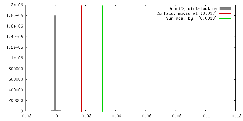
| Density |
| ||||||||||||||||||||||||||||||||||||||||||||||||||||||||||||||||||||
| Symmetry | Space group: 1 | ||||||||||||||||||||||||||||||||||||||||||||||||||||||||||||||||||||
| Details | EMDB XML:
CCP4 map header:
| ||||||||||||||||||||||||||||||||||||||||||||||||||||||||||||||||||||
 Z (Sec.)
 Y (Row.)
 X (Col.)


-Supplemental data
- Sample components
-Entire : murine p53
| Entire | Name: murine p53 |
|---|---|
| Components |
|
-Supramolecule #1000: murine p53
| Supramolecule | Name: murine p53 / type: sample / ID: 1000 / Details: homogenous sample / Oligomeric state: tetramer / Number unique components: 1 |
|---|---|
| Molecular weight | Experimental: 180 KDa / Theoretical: 174 KDa / Method: ultracentrifugation |
-Macromolecule #1: p53
| Macromolecule | Name: p53 / type: protein_or_peptide / ID: 1 / Name.synonym: tumour suppressor / Details: ATP-g-S complex / Number of copies: 4 / Oligomeric state: tetramer / Recombinant expression: Yes |
|---|---|
| Source (natural) | Organism: |
| Molecular weight | Experimental: 43 KDa / Theoretical: 45 KDa |
| Recombinant expression | Organism: Baculoviral system in Sf9 insect cells / Recombinant plasmid: pVL1393 |
| Sequence | GO: nucleus / InterPro: p53 tumour suppressor family |
-Experimental details
-Structure determination
| Method | cryo EM |
|---|---|
 Processing Processing | single particle reconstruction |
| Aggregation state | particle |
-Sample preparation
| Buffer | pH: 7.5 / Details: 150mM NaCl, 5mM MgCl2, 25mM Tris-HCl |
|---|---|
| Grid | Details: R2/2 quantifoilMicro |
| Vitrification | Cryogen name: ETHANE / Chamber humidity: 80 % / Chamber temperature: 80 K / Instrument: OTHER / Details: Vitrification instrument: Vitrobot / Method: automatic blot |
- Electron microscopy
| Microscope | FEI/PHILIPS CM300FEG/T |
|---|---|
| Temperature | Min: 80 K / Max: 90 K / Average: 80 K |
| Alignment procedure | Legacy - Astigmatism: 110 |
| Date | Jan 2, 2004 |
| Image recording | Category: FILM / Film or detector model: KODAK SO-163 FILM / Digitization - Scanner: ZEISS SCAI / Digitization - Sampling interval: 7 µm / Number real images: 16 / Average electron dose: 20 e/Å2 / Od range: 0.7 / Bits/pixel: 8 |
| Electron beam | Acceleration voltage: 300 kV / Electron source:  FIELD EMISSION GUN FIELD EMISSION GUN |
| Electron optics | Calibrated magnification: 45000 / Illumination mode: OTHER / Imaging mode: BRIGHT FIELD / Cs: 2 mm / Nominal defocus max: 3.2 µm / Nominal defocus min: 1.7 µm / Nominal magnification: 45000 |
| Sample stage | Specimen holder: Eucentric / Specimen holder model: GATAN LIQUID NITROGEN |
-Image processing
| CTF correction | Details: Phase correction |
|---|---|
| Final reconstruction | Applied symmetry - Point group: D2 (2x2 fold dihedral) / Algorithm: OTHER / Resolution.type: BY AUTHOR / Resolution: 13.7 Å / Resolution method: FSC 0.5 CUT-OFF / Software - Name: Imagic / Details: Final map was obtained from 240 best classes / Number images used: 7000 |
| Final two d classification | Number classes: 400 |
-Atomic model buiding 1
| Initial model | PDB ID: Chain - Chain ID: B |
|---|---|
| Software | Name: URO |
| Details | PDBEntryID_givenInChain. Protocol: Rigid body. other PDB entries used in addition: 1c26 and 1t4f |
| Refinement | Space: RECIPROCAL / Protocol: RIGID BODY FIT / Target criteria: Best correlation |