 Movie
Movie Controller

[English] 日本語
 Yorodumi
- EMDB-1119: Three-dimensional structure of the bacteriophage P22 tail machine. -
+ Open data

- Basic information
| Entry | Database: EMDB / ID: EMD-1119 | |||||||||
|---|---|---|---|---|---|---|---|---|---|---|
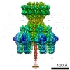
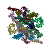
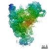
| Title | Three-dimensional structure of the bacteriophage P22 tail machine. | |||||||||
 Map data Map data | a cryoEM map of the P22 tail machine | |||||||||
 Sample Sample |
| |||||||||
| Biological species |  Enterobacteria phage P22 (virus) Enterobacteria phage P22 (virus) | |||||||||
| Method | single particle reconstruction / cryo EM / Resolution: 23.0 Å | |||||||||
 Authors Authors | Tang L / Marion WR / Cingolani G / Prevelige PE / Johnson JE | |||||||||
 Citation Citation | Journal: EMBO J / Year: 2005 Title: Three-dimensional structure of the bacteriophage P22 tail machine. Authors: Liang Tang / William R Marion / Gino Cingolani / Peter E Prevelige / John E Johnson /  Abstract: The tail of the bacteriophage P22 is composed of multiple protein components and integrates various biological functions that are crucial to the assembly and infection of the phage. The three- ...The tail of the bacteriophage P22 is composed of multiple protein components and integrates various biological functions that are crucial to the assembly and infection of the phage. The three-dimensional structure of the P22 tail machine determined by electron cryo-microscopy and image reconstruction reveals how the five types of polypeptides present as 51 subunits are organized into this molecular machine through twelve-, six- and three-fold symmetry, and provides insights into molecular events during host cell attachment and phage DNA translocation. | |||||||||
| History |
|
- Structure visualization
| Movie |
 Movie viewer Movie viewer |
|---|---|
| Structure viewer | EM map: SurfViewMolmilJmol/JSmol |
| Supplemental images |
- Downloads & links
-EMDB archive
| Map data | emd_1119.map.gz | 1.4 MB | EMDB map data format | |
|---|---|---|---|---|
| Header (meta data) | emd-1119-v30.xmlemd-1119.xml | 13.8 KB 13.8 KB | Display Display | EMDB header |
| Images |  1119.gif 1119.gif | 23.8 KB | ||
| Archive directory |  http://ftp.pdbj.org/pub/emdb/structures/EMD-1119ftp://ftp.pdbj.org/pub/emdb/structures/EMD-1119 http://ftp.pdbj.org/pub/emdb/structures/EMD-1119ftp://ftp.pdbj.org/pub/emdb/structures/EMD-1119 | HTTPS FTP |
-Related structure data
| Similar structure data |
|---|


-Links
| EMDB pages | EMDB (EBI/PDBe) / EMDataResource |
|---|
-Map
| File | Download / File: emd_1119.map.gz / Format: CCP4 / Size: 15.3 MB / Type: IMAGE STORED AS FLOATING POINT NUMBER (4 BYTES) | ||||||||||||||||||||||||||||||||||||||||||||||||||||||||||||||||||||
|---|---|---|---|---|---|---|---|---|---|---|---|---|---|---|---|---|---|---|---|---|---|---|---|---|---|---|---|---|---|---|---|---|---|---|---|---|---|---|---|---|---|---|---|---|---|---|---|---|---|---|---|---|---|---|---|---|---|---|---|---|---|---|---|---|---|---|---|---|---|
| Annotation | a cryoEM map of the P22 tail machine | ||||||||||||||||||||||||||||||||||||||||||||||||||||||||||||||||||||
| Projections & slices | Image control
Images are generated by Spider. | ||||||||||||||||||||||||||||||||||||||||||||||||||||||||||||||||||||
| Voxel size | X=Y=Z: 4.2 Å | ||||||||||||||||||||||||||||||||||||||||||||||||||||||||||||||||||||
| Density |
| ||||||||||||||||||||||||||||||||||||||||||||||||||||||||||||||||||||
| Symmetry | Space group: 1 | ||||||||||||||||||||||||||||||||||||||||||||||||||||||||||||||||||||
| Details | EMDB XML:
CCP4 map header:
| ||||||||||||||||||||||||||||||||||||||||||||||||||||||||||||||||||||
 Z (Sec.)
 Y (Row.)
 X (Col.)


-Supplemental data
- Sample components
-Entire : the tail machine isolated from bacteriophage P22
| Entire | Name: the tail machine isolated from bacteriophage P22 |
|---|---|
| Components |
|
-Supramolecule #1000: the tail machine isolated from bacteriophage P22
| Supramolecule | Name: the tail machine isolated from bacteriophage P22 / type: sample / ID: 1000 / Oligomeric state: 12-, 6-, and 3-fold symmetry / Number unique components: 5 |
|---|---|
| Molecular weight | Theoretical: 2.8 MDa |
-Macromolecule #1: portal
| Macromolecule | Name: portal / type: protein_or_peptide / ID: 1 / Name.synonym: gp1 / Details: DNA packaging / Number of copies: 12 / Oligomeric state: dodecamer / Recombinant expression: Yes |
|---|---|
| Source (natural) | Organism: Enterobacteria phage P22 (virus) / Strain: 13-am H101 |
| Molecular weight | Experimental: 82.611 MDa / Theoretical: 83 MDa |
| Recombinant expression | Organism: Salmonella enterica Serovar Typhimurium strain |
-Macromolecule #2: tailspike
| Macromolecule | Name: tailspike / type: protein_or_peptide / ID: 2 / Name.synonym: gp9 / Details: receptor binding / Number of copies: 18 / Oligomeric state: trimer / Recombinant expression: Yes |
|---|---|
| Source (natural) | Organism: Enterobacteria phage P22 (virus) / Strain: 13-am H101 |
| Molecular weight | Experimental: 71.857 MDa / Theoretical: 70 MDa |
| Recombinant expression | Organism: Salmonella enterica Serovar Typhimurium strain |
-Macromolecule #3: gp4
| Macromolecule | Name: gp4 / type: protein_or_peptide / ID: 3 / Name.synonym: accessory protein / Details: forms a channel; transglycosylase / Number of copies: 12 / Oligomeric state: dodecamer / Recombinant expression: Yes |
|---|---|
| Source (natural) | Organism: Enterobacteria phage P22 (virus) / Strain: 13-am H101 |
| Molecular weight | Experimental: 18.025 MDa / Theoretical: 20 MDa |
| Recombinant expression | Organism: Salmonella enterica Serovar Typhimurium strain |
-Macromolecule #4: gp10
| Macromolecule | Name: gp10 / type: protein_or_peptide / ID: 4 / Name.synonym: accessory protein / Details: forms a channel / Number of copies: 6 / Oligomeric state: hexamer / Recombinant expression: Yes |
|---|---|
| Source (natural) | Organism: Enterobacteria phage P22 (virus) / Strain: 13-am H101 |
| Molecular weight | Experimental: 52.457 MDa / Theoretical: 50 MDa |
| Recombinant expression | Organism: Salmonella enterica Serovar Typhimurium strain |
-Macromolecule #5: gp26
| Macromolecule | Name: gp26 / type: protein_or_peptide / ID: 5 / Name.synonym: accessory protein / Details: needle-like plug / Number of copies: 3 / Oligomeric state: trimer / Recombinant expression: Yes |
|---|---|
| Source (natural) | Organism: Enterobacteria phage P22 (virus) / Strain: 13-am H101 |
| Molecular weight | Experimental: 24.603 MDa / Theoretical: 25 MDa |
| Recombinant expression | Organism: Salmonella enterica Serovar Typhimurium strain |
-Experimental details
-Structure determination
| Method | cryo EM |
|---|---|
 Processing Processing | single particle reconstruction |
| Aggregation state | particle |
-Sample preparation
| Buffer | pH: 8 / Details: 50 mM Na2HPO4, pH8.0 |
|---|---|
| Grid | Details: 300 mesh holey copper grid |
| Vitrification | Cryogen name: ETHANE / Chamber temperature: 89 K / Instrument: HOMEMADE PLUNGER / Details: Vitrification instrument: home-made plunger / Method: blot for 3-4 seconds before plunging |
- Electron microscopy
| Microscope | FEI/PHILIPS CM200FEG |
|---|---|
| Temperature | Average: 89 K |
| Alignment procedure | Legacy - Astigmatism: objective lens astigmatism was corrected at 105,000 times magnification |
| Date | May 19, 2004 |
| Image recording | Category: FILM / Film or detector model: KODAK SO-163 FILM / Digitization - Scanner: ZEISS SCAI / Digitization - Sampling interval: 7 µm / Number real images: 54 / Bits/pixel: 8 |
| Tilt angle min | 0 |
| Electron beam | Acceleration voltage: 120 kV / Electron source:  FIELD EMISSION GUN FIELD EMISSION GUN |
| Electron optics | Illumination mode: OTHER / Imaging mode: BRIGHT FIELD / Cs: 2.0 mm / Nominal magnification: 50000 |
| Sample stage | Specimen holder: Side entry liquid nitrogen-cooled cryo specimen holder Specimen holder model: GATAN LIQUID NITROGEN / Tilt angle max: 60 |
-Image processing
| Details | The particles showed preferential orientation on the grids. CryoEM data were collected using the tilt method. The tilt angles ranged from 0-60 degrees. The particles were selected with a semi-automated selection program. |
|---|---|
| Final reconstruction | Applied symmetry - Point group: C6 (6 fold cyclic) / Algorithm: OTHER / Resolution.type: BY AUTHOR / Resolution: 23.0 Å / Resolution method: FSC 0.5 CUT-OFF / Software - Name: spider / Number images used: 11329 |
| Final angle assignment | Details: SPIDER:theta 90 degrees, phi 60 degrees |
-Atomic model buiding 1
| Initial model | (PDB ID: , ) |
|---|---|
| Software | Name: colores |
| Details | Protocol: rigid body. The receptor-binding domain (1TYX) of the tailspike was computationally docked with the program colores, and the docking was unambiguous. Then the head-binding domain (1LKT) was manually docked. |
| Refinement | Space: REAL / Protocol: RIGID BODY FIT / Target criteria: correlation coefficient |


