 Movie
Movie Controller

[English] 日本語
 Yorodumi
- EMDB-1118: Structural polymorphism of the major capsid protein of a double-s... -
+ Open data

- Basic information
| Entry | Database: EMDB / ID: EMD-1118 | |||||||||
|---|---|---|---|---|---|---|---|---|---|---|
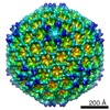
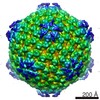
| Title | Structural polymorphism of the major capsid protein of a double-stranded RNA virus: an amphipathic alpha helix as a molecular switch. | |||||||||
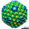
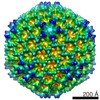
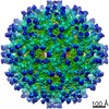
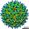
 Map data Map data | Three-dimensional density map of IBDV chimeric capsids at 15 A resolution, viewed along 2 fold axis of symmetry. The chimeric IBDV protein is VP2, including 466 amino acid residues,with an N-terminal His tag | |||||||||
 Sample Sample |
| |||||||||
| Biological species |  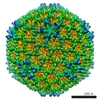 Infectious bursal disease virus (Gumboro virus) Infectious bursal disease virus (Gumboro virus) | |||||||||
| Method | single particle reconstruction / cryo EM / Resolution: 15.0 Å | |||||||||
 Authors Authors | Saugar I / Luque D / Ona A / Rodriguez JF / Carrascosa JL / Trus BL / Caston JR | |||||||||
 Citation Citation | Journal: Structure / Year: 2005 Title: Structural polymorphism of the major capsid protein of a double-stranded RNA virus: an amphipathic alpha helix as a molecular switch. Authors: Irene Saugar / Daniel Luque / Ana Oña / José F Rodríguez / José L Carrascosa / Benes L Trus / José R Castón /  Abstract: The infectious bursal disease virus T=13 viral particle is composed of two major proteins, VP2 and VP3. Here, we show that the molecular basis of the conformational flexibility of the major capsid ...The infectious bursal disease virus T=13 viral particle is composed of two major proteins, VP2 and VP3. Here, we show that the molecular basis of the conformational flexibility of the major capsid protein precursor, pVP2, is an amphipatic alpha helix formed by the sequence GFKDIIRAIR. VP2 containing this alpha helix is able to assemble into the T=13 capsid only when expressed as a chimeric protein with an N-terminal His tag. An amphiphilic alpha helix, which acts as a conformational switch, is thus responsible for the inherent structural polymorphism of VP2. The His tag mimics the VP3 C-terminal region closely and acts as a molecular triggering factor. Using cryo-electron microscopy difference imaging, both polypeptide elements were detected on the capsid inner surface. We propose that electrostatic interactions between these two morphogenic elements are transmitted to VP2 to acquire the competent conformations for capsid assembly. | |||||||||
| History |
|
- Structure visualization
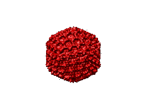
| Movie |
 Movie viewer Movie viewer |
|---|---|
| Structure viewer | EM map: SurfViewMolmilJmol/JSmol |
| Supplemental images |
- Downloads & links
-EMDB archive
| Map data | emd_1118.map.gz | 4.8 MB | EMDB map data format | |
|---|---|---|---|---|
| Header (meta data) | emd-1118-v30.xmlemd-1118.xml | 9.4 KB 9.4 KB | Display Display | EMDB header |
| Images |  1118.gif 1118.gif | 11.6 KB | ||
| Archive directory |  http://ftp.pdbj.org/pub/emdb/structures/EMD-1118ftp://ftp.pdbj.org/pub/emdb/structures/EMD-1118 http://ftp.pdbj.org/pub/emdb/structures/EMD-1118ftp://ftp.pdbj.org/pub/emdb/structures/EMD-1118 | HTTPS FTP |
-Related structure data


-Links
| EMDB pages | EMDB (EBI/PDBe) / EMDataResource |
|---|
-Map
| File | Download / File: emd_1118.map.gz / Format: CCP4 / Size: 25.2 MB / Type: IMAGE STORED AS FLOATING POINT NUMBER (4 BYTES) | ||||||||||||||||||||||||||||||||||||||||||||||||||||||||||||||||||||
|---|---|---|---|---|---|---|---|---|---|---|---|---|---|---|---|---|---|---|---|---|---|---|---|---|---|---|---|---|---|---|---|---|---|---|---|---|---|---|---|---|---|---|---|---|---|---|---|---|---|---|---|---|---|---|---|---|---|---|---|---|---|---|---|---|---|---|---|---|---|
| Annotation | Three-dimensional density map of IBDV chimeric capsids at 15 A resolution, viewed along 2 fold axis of symmetry. The chimeric IBDV protein is VP2, including 466 amino acid residues,with an N-terminal His tag | ||||||||||||||||||||||||||||||||||||||||||||||||||||||||||||||||||||
| Projections & slices | Image control
Images are generated by Spider. | ||||||||||||||||||||||||||||||||||||||||||||||||||||||||||||||||||||
| Voxel size | X=Y=Z: 4.2 Å | ||||||||||||||||||||||||||||||||||||||||||||||||||||||||||||||||||||
| Density |
| ||||||||||||||||||||||||||||||||||||||||||||||||||||||||||||||||||||
| Symmetry | Space group: 1 | ||||||||||||||||||||||||||||||||||||||||||||||||||||||||||||||||||||
| Details | EMDB XML:
CCP4 map header:
| ||||||||||||||||||||||||||||||||||||||||||||||||||||||||||||||||||||
 Z (Sec.)
 Y (Row.)
 X (Col.)


-Supplemental data
- Sample components
-Entire : chimeric Infectious Bursal Disease Virus capsid
| Entire | Name: chimeric Infectious Bursal Disease Virus capsid |
|---|---|
| Components |
|
-Supramolecule #1000: chimeric Infectious Bursal Disease Virus capsid
| Supramolecule | Name: chimeric Infectious Bursal Disease Virus capsid / type: sample / ID: 1000 / Number unique components: 1 |
|---|
-Supramolecule #1: Infectious bursal disease virus
| Supramolecule | Name: Infectious bursal disease virus / type: virus / ID: 1 / Name.synonym: HT-VP2-466 / NCBI-ID: 10995 / Sci species name: Infectious bursal disease virus / Virus type: VIRUS-LIKE PARTICLE / Virus isolate: STRAIN / Virus enveloped: No / Virus empty: Yes / Syn species name: HT-VP2-466 |
|---|---|
| Host (natural) | Organism:  |
| Virus shell | Shell ID: 1 / Name: HT-VP2-466 single layered capsid / Diameter: 700 Å / T number (triangulation number): 13 |
-Experimental details
-Structure determination
| Method | cryo EM |
|---|---|
 Processing Processing | single particle reconstruction |
| Aggregation state | particle |
-Sample preparation
| Concentration | 2 mg/mL |
|---|---|
| Buffer | pH: 6.2 / Details: PIPES 25 mM, 150 mM NaCl, 20 mM CaCl2 |
| Grid | Details: Holey carbon film on copper grid |
| Vitrification | Cryogen name: ETHANE / Chamber temperature: 111 K / Instrument: HOMEMADE PLUNGER / Details: Vitrification instrument: manual / Method: double blotting |
- Electron microscopy
| Microscope | FEI TECNAI 20 |
|---|---|
| Temperature | Average: 98 K |
| Alignment procedure | Legacy - Astigmatism: objective lens astigmatism was corrected at 150,000 times |
| Details | TEM Tecnai G2 |
| Image recording | Category: FILM / Film or detector model: KODAK SO-163 FILM / Digitization - Scanner: ZEISS SCAI / Digitization - Sampling interval: 7 µm / Number real images: 82 / Average electron dose: 9 e/Å2 / Details: original images were binned 3X / Od range: 1 / Bits/pixel: 8 |
| Electron beam | Acceleration voltage: 200 kV / Electron source:  FIELD EMISSION GUN FIELD EMISSION GUN |
| Electron optics | Calibrated magnification: 49700 / Illumination mode: FLOOD BEAM / Imaging mode: BRIGHT FIELD / Cs: 2.26 mm / Nominal defocus max: 3.0 µm / Nominal defocus min: 0.3 µm / Nominal magnification: 50000 |
| Sample stage | Specimen holder: Gatan / Specimen holder model: GATAN LIQUID NITROGEN |
-Image processing
| CTF correction | Details: Each particle |
|---|---|
| Final reconstruction | Applied symmetry - Point group: I (icosahedral) / Algorithm: OTHER / Resolution.type: BY AUTHOR / Resolution: 15.0 Å / Resolution method: FSC 0.5 CUT-OFF / Software - Name: emPFT / Number images used: 1557 |
