 ムービー
ムービー コントローラー


+ データを開く

- 基本情報
基本情報
| 登録情報 | データベース: EMDB / ID: EMD-11057 | ||||||||||||
|---|---|---|---|---|---|---|---|---|---|---|---|---|---|
| タイトル | Cryo-electron tomogram after FIB-milling of Planctomycetes species Tuwongella immobilis (#2). | ||||||||||||
 マップデータ マップデータ | Whole-cell cryo electron tomogram after FIB-milling of Tuwongella immobilis | ||||||||||||
 試料 試料 |
| ||||||||||||
| 生物種 |  Tuwongella immobilis (バクテリア) Tuwongella immobilis (バクテリア) | ||||||||||||
| 手法 | 電子線トモグラフィー法 / クライオ電子顕微鏡法 | ||||||||||||
 データ登録者 データ登録者 | Seeger C / Andersson SGE | ||||||||||||
| 資金援助 |  スウェーデン, 3件 スウェーデン, 3件
| ||||||||||||
 引用 引用 | ジャーナル: Genome Biol Evol / 年: 2020 タイトル: Evolutionary Remodeling of the Cell Envelope in Bacteria of the Planctomycetes Phylum. 著者: Mayank Mahajan / Christian Seeger / Benjamin Yee / Siv G E Andersson / 要旨: Bacteria of the Planctomycetes phylum have many unique cellular features, such as extensive membrane invaginations and the ability to import macromolecules. These features raise intriguing questions ...Bacteria of the Planctomycetes phylum have many unique cellular features, such as extensive membrane invaginations and the ability to import macromolecules. These features raise intriguing questions about the composition of their cell envelopes. In this study, we have used microscopy, phylogenomics, and proteomics to examine the composition and evolution of cell envelope proteins in Tuwongella immobilis and other members of the Planctomycetes. Cryo-electron tomography data indicated a distance of 45 nm between the inner and outer membranes in T. immobilis. Consistent with the wide periplasmic space, our bioinformatics studies showed that the periplasmic segments of outer-membrane proteins in type II secretion systems are extended in bacteria of the order Planctomycetales. Homologs of two highly abundant cysteine-rich cell wall proteins in T. immobilis were identified in all members of the Planctomycetales, whereas genes for peptidoglycan biosynthesis and cell elongation have been lost in many members of this bacterial group. The cell wall proteins contain multiple copies of the YTV motif, which is the only domain that is conserved and unique to the Planctomycetales. Earlier diverging taxa in the Planctomycetes phylum contain genes for peptidoglycan biosynthesis but no homologs to the YTV cell wall proteins. The major remodeling of the cell envelope in the ancestor of the Planctomycetales coincided with the emergence of budding and other unique cellular phenotypes. The results have implications for hypotheses about the process whereby complex cellular features evolve in bacteria. | ||||||||||||
| 履歴 |
|
- 構造の表示
構造の表示
| ムービー |
 ムービービューア ムービービューア |
|---|---|
| 添付画像 |

- ダウンロードとリンク
-EMDBアーカイブ
| マップデータ | emd_11057.map.gz | 1.1 GB | EMDBマップデータ形式 | |
|---|---|---|---|---|
| ヘッダ (付随情報) | emd-11057-v30.xmlemd-11057.xml | 12.9 KB 12.9 KB | 表示 表示 | EMDBヘッダ |
| 画像 | 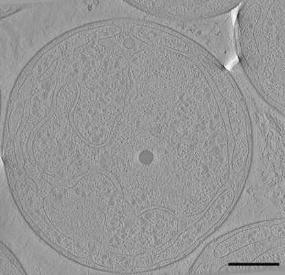 emd_11057.png emd_11057.png | 127 KB | ||
| アーカイブディレクトリ |  http://ftp.pdbj.org/pub/emdb/structures/EMD-11057ftp://ftp.pdbj.org/pub/emdb/structures/EMD-11057 http://ftp.pdbj.org/pub/emdb/structures/EMD-11057ftp://ftp.pdbj.org/pub/emdb/structures/EMD-11057 | HTTPS FTP |
-検証レポート
| 文書・要旨 | emd_11057_validation.pdf.gz | 142.5 KB | 表示 | EMDB検証レポート |
|---|---|---|---|---|
| 文書・詳細版 | emd_11057_full_validation.pdf.gz | 141.6 KB | 表示 | |
| XML形式データ | emd_11057_validation.xml.gz | 3.9 KB | 表示 | |
| アーカイブディレクトリ | https://ftp.pdbj.org/pub/emdb/validation_reports/EMD-11057ftp://ftp.pdbj.org/pub/emdb/validation_reports/EMD-11057 | HTTPS FTP |
-関連構造データ
| 関連構造データ | C: 同じ文献を引用 ( |
|---|---|
| 電子顕微鏡画像生データ | EMPIAR-10448 (タイトル: Cryo electron tomography after FIB-milling of Planctomycetes species Tuwongella immobilis Data size: 3.2 Data #1: Cryo-ET tilt series of Tuwongella immobilis after FIB-milling ("#2") [tilt series]) |
-リンク
| EMDBのページ | EMDB (EBI/PDBe) / EMDataResource |
|---|
-マップ
| ファイル | ダウンロード / ファイル: emd_11057.map.gz / 形式: CCP4 / 大きさ: 3.3 GB / タイプ: IMAGE STORED AS SIGNED BYTE | ||||||||||||||||||||||||||||||||||||||||||||||||||||||||||||
|---|---|---|---|---|---|---|---|---|---|---|---|---|---|---|---|---|---|---|---|---|---|---|---|---|---|---|---|---|---|---|---|---|---|---|---|---|---|---|---|---|---|---|---|---|---|---|---|---|---|---|---|---|---|---|---|---|---|---|---|---|---|
| 注釈 | Whole-cell cryo electron tomogram after FIB-milling of Tuwongella immobilis | ||||||||||||||||||||||||||||||||||||||||||||||||||||||||||||
| ボクセルのサイズ | X=Y=Z: 7.286 Å | ||||||||||||||||||||||||||||||||||||||||||||||||||||||||||||
| 密度 |
| ||||||||||||||||||||||||||||||||||||||||||||||||||||||||||||
| 対称性 | 空間群: 1 | ||||||||||||||||||||||||||||||||||||||||||||||||||||||||||||
| 詳細 | EMDB XML:
CCP4マップ ヘッダ情報:
| ||||||||||||||||||||||||||||||||||||||||||||||||||||||||||||
-添付データ
- 試料の構成要素
-全体 : Tuwongella immobilis, whole cell cryo-electron tomogram after FIB...
| 全体 | 名称: Tuwongella immobilis, whole cell cryo-electron tomogram after FIB-milling. |
|---|---|
| 要素 |
|
-超分子 #1: Tuwongella immobilis, whole cell cryo-electron tomogram after FIB...
| 超分子 | 名称: Tuwongella immobilis, whole cell cryo-electron tomogram after FIB-milling. タイプ: cell / ID: 1 / 親要素: 0 詳細: Whole cell tomogram of Tuwongella immobilis revealing the invaginated cytoplasmic membrane, which is characteristic for many members of the phylum Planctomycetes. Trans-membrane protein ...詳細: Whole cell tomogram of Tuwongella immobilis revealing the invaginated cytoplasmic membrane, which is characteristic for many members of the phylum Planctomycetes. Trans-membrane protein complexes, like pili and other secretion systems are spanning the cell envelope. The ribosome-rich compartment corresponds to the cytoplasm, the ribosome-free compartment corresponds to the enlarged periplasm. |
|---|---|
| 由来(天然) | 生物種: Tuwongella immobilis (バクテリア) |
-実験情報
-構造解析
| 手法 | クライオ電子顕微鏡法 |
|---|---|
 解析 解析 | 電子線トモグラフィー法 |
| 試料の集合状態 | cell |
-試料調製
| 緩衝液 | pH: 7.4 / 構成要素 - 濃度: 10.0 mmol/L / 構成要素 - 名称: Na-phosphate 詳細: 10 mM Na-phosphate buffer, pH 7.4, 0.2 microm filtered. DO NOT use salt or any other components that increase osmolarity! |
|---|---|
| グリッド | モデル: Quantifoil R2/1 / 材質: GOLD / メッシュ: 200 / 支持フィルム - 材質: CARBON / 支持フィルム - トポロジー: HOLEY / 前処理 - タイプ: GLOW DISCHARGE / 前処理 - 雰囲気: AIR / 前処理 - 気圧: 101.325 kPa |
| 凍結 | 凍結剤: ETHANE / チャンバー内湿度: 100 % / チャンバー内温度: 293 K / 装置: FEI VITROBOT MARK IV 詳細: Blot time: 6s, blot force: -5, wait time: 15s, 293K, 100% humidity. |
| 詳細 | Bacterial cells grown on M1 agar plates at 32degrees celsius and kept at 20degress celsius until vitrification |
| Cryo protectant | No |
| 切片作成 | その他: NO SECTIONING |
- 電子顕微鏡法
| 顕微鏡 | FEI TITAN KRIOS |
|---|---|
| 温度 | 最低: 93.0 K |
| 特殊光学系 | エネルギーフィルター - スリット幅: 20 eV |
| 詳細 | Before tilt series acquisition, cryo focused ion beam (FIB) milling with gallium ion source was performed on a FEI Dual beam Scios (Thermo Fisher Scientific, Netherlands). Autogrids with vitrified cells were mounted onto a dedicated cryo holder and transferred onto a cooled (93K) cryo stage in the dual beam microscope. The lamellae were prepared with two parallel rectangular patterns at both sides (top and bottom) of the cells, at milling angle of 10-15deg, accelerating voltage of 30 kV and ion currents between 30-300 pA. The final thickness of the lamella (milled at the lowest ion current) was about 150-300 nm. |
| 撮影 | フィルム・検出器のモデル: GATAN K2 SUMMIT (4k x 4k) 実像数: 121 / 平均電子線量: 0.82 e/Å2 詳細: Total dose tilt series: 99 electrons/angstrom; number of tilts: 121; dose per tilt: 0.82 electron/angstrom |
| 電子線 | 加速電圧: 300 kV / 電子線源:  FIELD EMISSION GUN FIELD EMISSION GUN |
| 電子光学系 | C2レンズ絞り径: 70.0 µm / 倍率(補正後): 19500 / 照射モード: OTHER / 撮影モード: OTHER / 倍率(公称値): 19500 |
| 試料ステージ | 試料ホルダーモデル: FEI TITAN KRIOS AUTOGRID HOLDER ホルダー冷却材: NITROGEN |
| 実験機器 |  モデル: Titan Krios / 画像提供: FEI Company |
-画像解析
| 最終 再構成 | アルゴリズム: BACK PROJECTION / ソフトウェア - 名称: IMOD (ver. 4.9.10) / 使用した粒子像数: 121 |
|---|
