 Movie
Movie Controller

[English] 日本語
 Yorodumi
- EMDB-11074: Cryo-electron tomogram after FIB-milling of Gemmata obscuriglobus... -
+ Open data

- Basic information
| Entry | Database: EMDB / ID: EMD-11074 | ||||||||||||
|---|---|---|---|---|---|---|---|---|---|---|---|---|---|
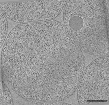
| Title | Cryo-electron tomogram after FIB-milling of Gemmata obscuriglobus (#1), a species of the Planctomycetes phylum. | ||||||||||||
 Map data Map data | Cryo-electron tomogram after FIB-milling of Gemmata obscuriglobus, a species of the Planctomycetes phylum | ||||||||||||
 Sample Sample |
| ||||||||||||
| Biological species |  Gemmata obscurig (bacteria) Gemmata obscurig (bacteria) | ||||||||||||
| Method | electron tomography / cryo EM | ||||||||||||
 Authors Authors | Seeger C / Andersson SGE | ||||||||||||
| Funding support |  Sweden, 3 items Sweden, 3 items
| ||||||||||||
 Citation Citation | Journal: Genome Biol Evol / Year: 2020 Title: Evolutionary Remodeling of the Cell Envelope in Bacteria of the Planctomycetes Phylum. Authors: Mayank Mahajan / Christian Seeger / Benjamin Yee / Siv G E Andersson / Abstract: Bacteria of the Planctomycetes phylum have many unique cellular features, such as extensive membrane invaginations and the ability to import macromolecules. These features raise intriguing questions ...Bacteria of the Planctomycetes phylum have many unique cellular features, such as extensive membrane invaginations and the ability to import macromolecules. These features raise intriguing questions about the composition of their cell envelopes. In this study, we have used microscopy, phylogenomics, and proteomics to examine the composition and evolution of cell envelope proteins in Tuwongella immobilis and other members of the Planctomycetes. Cryo-electron tomography data indicated a distance of 45 nm between the inner and outer membranes in T. immobilis. Consistent with the wide periplasmic space, our bioinformatics studies showed that the periplasmic segments of outer-membrane proteins in type II secretion systems are extended in bacteria of the order Planctomycetales. Homologs of two highly abundant cysteine-rich cell wall proteins in T. immobilis were identified in all members of the Planctomycetales, whereas genes for peptidoglycan biosynthesis and cell elongation have been lost in many members of this bacterial group. The cell wall proteins contain multiple copies of the YTV motif, which is the only domain that is conserved and unique to the Planctomycetales. Earlier diverging taxa in the Planctomycetes phylum contain genes for peptidoglycan biosynthesis but no homologs to the YTV cell wall proteins. The major remodeling of the cell envelope in the ancestor of the Planctomycetales coincided with the emergence of budding and other unique cellular phenotypes. The results have implications for hypotheses about the process whereby complex cellular features evolve in bacteria. | ||||||||||||
| History |
|
- Structure visualization
| Movie |
 Movie viewer Movie viewer |
|---|---|
| Supplemental images |
- Downloads & links
-EMDB archive
| Map data | emd_11074.map.gz | 758.5 MB | EMDB map data format | |
|---|---|---|---|---|
| Header (meta data) | emd-11074-v30.xmlemd-11074.xml | 12.9 KB 12.9 KB | Display Display | EMDB header |
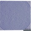
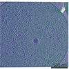
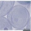
| Images |  emd_11074.png emd_11074.png | 126.1 KB | ||
| Archive directory |  http://ftp.pdbj.org/pub/emdb/structures/EMD-11074ftp://ftp.pdbj.org/pub/emdb/structures/EMD-11074 http://ftp.pdbj.org/pub/emdb/structures/EMD-11074ftp://ftp.pdbj.org/pub/emdb/structures/EMD-11074 | HTTPS FTP |
-Related structure data
| Related structure data | C: citing same article ( |
|---|---|
| EM raw data | EMPIAR-10449 (Title: Cryo electron tomography after FIB-milling of Planctomycetes species Gemmata obscuriglobus (#1) Data size: 2.7 Data #1: Cryo-ET tilt series of Gemmata obscuriglobus after FIB-milling (#1/2) [tilt series]) |
-Links
| EMDB pages | EMDB (EBI/PDBe) / EMDataResource |
|---|
-Map
| File | Download / File: emd_11074.map.gz / Format: CCP4 / Size: 2.3 GB / Type: IMAGE STORED AS SIGNED BYTE | ||||||||||||||||||||||||||||||||||||||||||||||||||||||||||||
|---|---|---|---|---|---|---|---|---|---|---|---|---|---|---|---|---|---|---|---|---|---|---|---|---|---|---|---|---|---|---|---|---|---|---|---|---|---|---|---|---|---|---|---|---|---|---|---|---|---|---|---|---|---|---|---|---|---|---|---|---|---|
| Annotation | Cryo-electron tomogram after FIB-milling of Gemmata obscuriglobus, a species of the Planctomycetes phylum | ||||||||||||||||||||||||||||||||||||||||||||||||||||||||||||
| Voxel size | X=Y=Z: 7.286 Å | ||||||||||||||||||||||||||||||||||||||||||||||||||||||||||||
| Density |
| ||||||||||||||||||||||||||||||||||||||||||||||||||||||||||||
| Symmetry | Space group: 1 | ||||||||||||||||||||||||||||||||||||||||||||||||||||||||||||
| Details | EMDB XML:
CCP4 map header:
| ||||||||||||||||||||||||||||||||||||||||||||||||||||||||||||
-Supplemental data
- Sample components
-Entire : Gemmata obscuriglobus, whole cell cryo-electron tomogram after FI...
| Entire | Name: Gemmata obscuriglobus, whole cell cryo-electron tomogram after FIB-milling. |
|---|---|
| Components |
|
-Supramolecule #1: Gemmata obscuriglobus, whole cell cryo-electron tomogram after FI...
| Supramolecule | Name: Gemmata obscuriglobus, whole cell cryo-electron tomogram after FIB-milling. type: cell / ID: 1 / Parent: 0 Details: Whole cell tomogram of Gemmata obscuriglobus revealing the invaginated cytoplasmic membrane, which is characteristic for many members of the phylum Planctomycetes. The ribosome-rich ...Details: Whole cell tomogram of Gemmata obscuriglobus revealing the invaginated cytoplasmic membrane, which is characteristic for many members of the phylum Planctomycetes. The ribosome-rich cytoplasm is separated by the invaginated cytoplasmic membrane from the enlarged periplasmic space. |
|---|---|
| Source (natural) | Organism: Gemmata obscurig (bacteria) |
-Experimental details
-Structure determination
| Method | cryo EM |
|---|---|
 Processing Processing | electron tomography |
| Aggregation state | cell |
-Sample preparation
| Buffer | pH: 7.4 / Component - Concentration: 10.0 mmol/L / Component - Name: Na-phosphate Details: 10 mM Na-phosphate buffer, pH 7.4, 0.2 microm filtered. DO NOT use salt or any other components that increase osmolarity! |
|---|---|
| Grid | Model: Quantifoil R2/1 / Material: GOLD / Mesh: 200 / Support film - Material: CARBON / Support film - topology: HOLEY / Pretreatment - Type: GLOW DISCHARGE / Pretreatment - Atmosphere: AIR / Pretreatment - Pressure: 101.325 kPa |
| Vitrification | Cryogen name: ETHANE / Chamber humidity: 100 % / Chamber temperature: 293 K / Instrument: FEI VITROBOT MARK IV Details: Blot time: 6s, blot force: -5, wait time: 15s, 293K, 100% humidity. |
| Details | Bacterial cells grown on M1 agar plates at 32degrees celsius and kept at 20degress celsius until vitrification |
| Cryo protectant | No |
| Sectioning | Other: NO SECTIONING |
- Electron microscopy
| Microscope | FEI TITAN KRIOS |
|---|---|
| Temperature | Min: 93.0 K |
| Specialist optics | Energy filter - Slit width: 20 eV |
| Details | Before tilt series acquisition, cryo focused ion beam (FIB) milling with gallium ion source was performed on a FEI Dual beam Scios (Thermo Fisher Scientific, Netherlands). Autogrids with vitrified cells were mounted onto a dedicated cryo holder and transferred onto a cooled (93K) cryo stage in the dual beam microscope. The lamellae were prepared with two parallel rectangular patterns at both sides (top and bottom) of the cells, at milling angle of 10-15deg, accelerating voltage of 30 kV and ion currents between 30-300 pA. The final thickness of the lamella (milled at the lowest ion current) was about 150-300 nm. |
| Image recording | Film or detector model: GATAN K2 SUMMIT (4k x 4k) / Number real images: 101 / Average electron dose: 1.0 e/Å2 Details: Total dose tilt series: 94.7 electrons/angstrom; number of tilts: 101; dose per tilt: approx. 1.0 electron/angstrom |
| Electron beam | Acceleration voltage: 300 kV / Electron source:  FIELD EMISSION GUN FIELD EMISSION GUN |
| Electron optics | C2 aperture diameter: 70.0 µm / Calibrated magnification: 19500 / Illumination mode: OTHER / Imaging mode: OTHER / Nominal magnification: 19500 |
| Sample stage | Specimen holder model: FEI TITAN KRIOS AUTOGRID HOLDER / Cooling holder cryogen: NITROGEN |
| Experimental equipment |  Model: Titan Krios / Image courtesy: FEI Company |
-Image processing
| Final reconstruction | Algorithm: BACK PROJECTION / Software - Name: IMOD (ver. 4.9.10) / Number images used: 101 |
|---|
