 Movie
Movie Controller

[English] 日本語
 Yorodumi
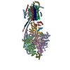
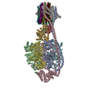
- EMDB-10998: Mitochondrial ATP synthase monomer from Rattus norvegicus domestica -
+ Open data

- Basic information
| Entry | Database: EMDB / ID: EMD-10998 | |||||||||
|---|---|---|---|---|---|---|---|---|---|---|
| Title | Mitochondrial ATP synthase monomer from Rattus norvegicus domestica | |||||||||
 Map data Map data | ||||||||||
 Sample Sample |
| |||||||||
| Biological species |  | |||||||||
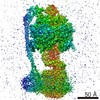
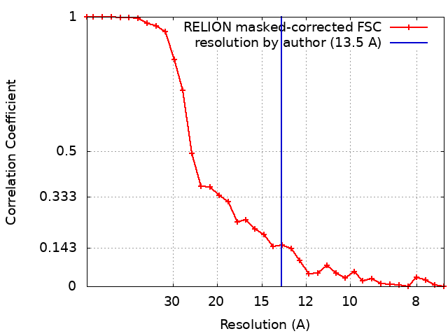
| Method | subtomogram averaging / cryo EM / Resolution: 13.5 Å | |||||||||
 Authors Authors | Chesnokov YM / Kamyshinsky RA | |||||||||
 Citation Citation | Journal: Nanotechnol Russia / Year: 2020 Title: Determining the Structure and Location of the ATP Synthase in the Membranes of Rat's Heart Mitochondria Using Cryoelectron Tomography Authors: Nesterov SV / Chesnokov YM / Kamyshinsky RA / Yaguzhinsky LS / Vasilov RG | |||||||||
| History |
|
- Structure visualization
| Movie |
 Movie viewer Movie viewer |
|---|---|
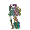
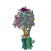
| Structure viewer | EM map: SurfViewMolmilJmol/JSmol |
| Supplemental images |

- Downloads & links
-EMDB archive
| Map data | emd_10998.map.gz | 682.9 KB | EMDB map data format | |
|---|---|---|---|---|
| Header (meta data) | emd-10998-v30.xmlemd-10998.xml | 16.1 KB 16.1 KB | Display Display | EMDB header |
| FSC (resolution estimation) | emd_10998_fsc.xml | 3 KB | Display | FSC data file |
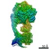
| Images | 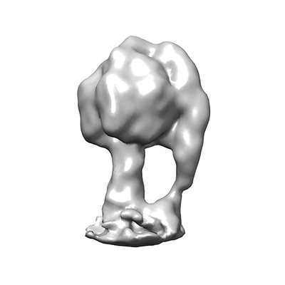 emd_10998.png emd_10998.png | 34.9 KB | ||
| Masks | emd_10998_msk_1.map | 2 MB | Mask map | |
| Others | emd_10998_half_map_1.map.gzemd_10998_half_map_2.map.gz | 1.4 MB 1.4 MB | ||
| Archive directory |  http://ftp.pdbj.org/pub/emdb/structures/EMD-10998ftp://ftp.pdbj.org/pub/emdb/structures/EMD-10998 http://ftp.pdbj.org/pub/emdb/structures/EMD-10998ftp://ftp.pdbj.org/pub/emdb/structures/EMD-10998 | HTTPS FTP |
-Validation report
| Summary document | emd_10998_validation.pdf.gz | 314.9 KB | Display | EMDB validaton report |
|---|---|---|---|---|
| Full document | emd_10998_full_validation.pdf.gz | 314.1 KB | Display | |
| Data in XML | emd_10998_validation.xml.gz | 8.5 KB | Display | |
| Arichive directory | https://ftp.pdbj.org/pub/emdb/validation_reports/EMD-10998ftp://ftp.pdbj.org/pub/emdb/validation_reports/EMD-10998 | HTTPS FTP |
-Related structure data
| Related structure data |  10997 |
|---|---|
| Similar structure data |


-Links
| EMDB pages | EMDB (EBI/PDBe) / EMDataResource |
|---|
-Map
| File | Download / File: emd_10998.map.gz / Format: CCP4 / Size: 2 MB / Type: IMAGE STORED AS FLOATING POINT NUMBER (4 BYTES) | ||||||||||||||||||||||||||||||||||||||||||||||||||||||||||||
|---|---|---|---|---|---|---|---|---|---|---|---|---|---|---|---|---|---|---|---|---|---|---|---|---|---|---|---|---|---|---|---|---|---|---|---|---|---|---|---|---|---|---|---|---|---|---|---|---|---|---|---|---|---|---|---|---|---|---|---|---|---|
| Projections & slices | Image control
Images are generated by Spider. | ||||||||||||||||||||||||||||||||||||||||||||||||||||||||||||
| Voxel size | X=Y=Z: 3.7 Å | ||||||||||||||||||||||||||||||||||||||||||||||||||||||||||||
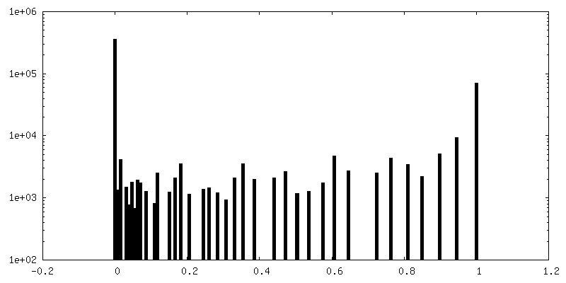
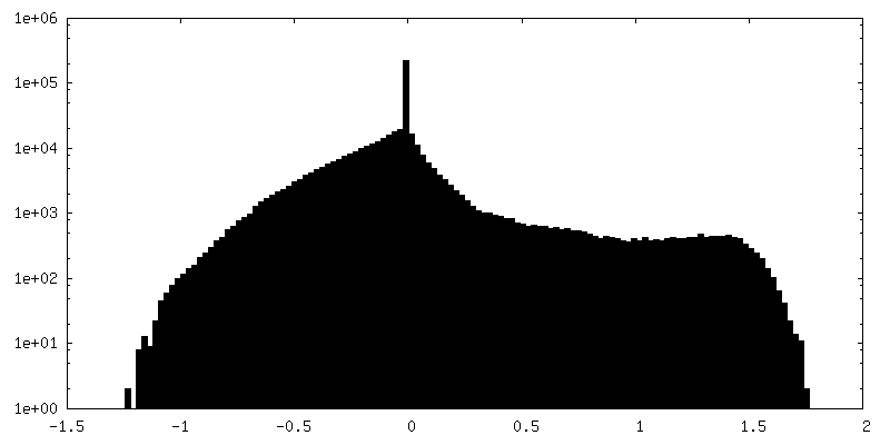
| Density |
| ||||||||||||||||||||||||||||||||||||||||||||||||||||||||||||
| Symmetry | Space group: 1 | ||||||||||||||||||||||||||||||||||||||||||||||||||||||||||||
| Details | EMDB XML:
CCP4 map header:
| ||||||||||||||||||||||||||||||||||||||||||||||||||||||||||||
 Z (Sec.)
 Y (Row.)
 X (Col.)


-Supplemental data
-Mask #1
| File | emd_10998_msk_1.map | ||||||||||||
|---|---|---|---|---|---|---|---|---|---|---|---|---|---|
| Projections & Slices |
| ||||||||||||
| Density Histograms |


-Half map: #2
| File | emd_10998_half_map_1.map | ||||||||||||
|---|---|---|---|---|---|---|---|---|---|---|---|---|---|
| Projections & Slices |
| ||||||||||||
| Density Histograms |


-Half map: #1
| File | emd_10998_half_map_2.map | ||||||||||||
|---|---|---|---|---|---|---|---|---|---|---|---|---|---|
| Projections & Slices |
| ||||||||||||
| Density Histograms |


- Sample components
-Entire : Mitochondrial ATP synthase dimer from Rattus norvegicus
| Entire | Name: Mitochondrial ATP synthase dimer from Rattus norvegicus |
|---|---|
| Components |
|
-Supramolecule #1: Mitochondrial ATP synthase dimer from Rattus norvegicus
| Supramolecule | Name: Mitochondrial ATP synthase dimer from Rattus norvegicus type: organelle_or_cellular_component / ID: 1 / Parent: 0 |
|---|---|
| Source (natural) | Organism: |
| Molecular weight | Theoretical: 600 KDa |
-Experimental details
-Structure determination
| Method | cryo EM |
|---|---|
 Processing Processing | subtomogram averaging |
| Aggregation state | 3D array |
-Sample preparation
| Concentration | 0.3 mg/mL |
|---|---|
| Buffer | pH: 7.4 |
| Grid | Model: Homemade / Material: COPPER / Mesh: 300 / Support film - Material: CARBON / Support film - topology: LACEY |
| Vitrification | Cryogen name: ETHANE / Chamber humidity: 100 % / Chamber temperature: 277 K / Instrument: FEI VITROBOT MARK IV / Details: blot for 2.5 seconds. |
- Electron microscopy
| Microscope | FEI TITAN KRIOS |
|---|---|
| Specialist optics | Spherical aberration corrector: Cs image corrector (CEOS, Germany) |
| Image recording | Film or detector model: FEI FALCON II (4k x 4k) / Detector mode: INTEGRATING / Digitization - Dimensions - Width: 4096 pixel / Digitization - Dimensions - Height: 4096 pixel / Digitization - Sampling interval: 14.0 µm / Number grids imaged: 1 / Average exposure time: 1.5 sec. / Average electron dose: 1.5 e/Å2 |
| Electron beam | Acceleration voltage: 300 kV / Electron source:  FIELD EMISSION GUN FIELD EMISSION GUN |
| Electron optics | C2 aperture diameter: 100.0 µm / Calibrated magnification: 37837 / Illumination mode: FLOOD BEAM / Imaging mode: BRIGHT FIELD / Cs: 0.01 mm / Nominal defocus max: 8.0 µm / Nominal defocus min: 6.0 µm / Nominal magnification: 18000 |
| Sample stage | Specimen holder model: FEI TITAN KRIOS AUTOGRID HOLDER / Cooling holder cryogen: NITROGEN |
| Experimental equipment |  Model: Titan Krios / Image courtesy: FEI Company |
+Image processing
-Atomic model buiding 1
| Initial model | PDB ID: |
|---|---|
| Refinement | Space: REAL / Protocol: RIGID BODY FIT / Target criteria: Correlation coefficient |

