Movie
Movie Controller


+ Open data

- Basic information
| Entry | Database: EMDB / ID: EMD-1088 | |||||||||
|---|---|---|---|---|---|---|---|---|---|---|
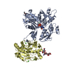
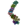
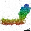
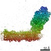
| Title | Structure of the acrosomal bundle. | |||||||||
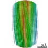
 Map data Map data | One unit cell of the acrosomal bundle, pseudo-centered to provide a orthogonal map from the pseudo-hexagonal packing | |||||||||
 Sample Sample |
| |||||||||
| Function / homology |  Function and homology information Function and homology informationcytoskeletal motor activator activity / myosin heavy chain binding / tropomyosin binding / actin filament bundle / troponin I binding / filamentous actin / mesenchyme migration / actin filament bundle assembly / skeletal muscle myofibril / striated muscle thin filament ...cytoskeletal motor activator activity / myosin heavy chain binding / tropomyosin binding / actin filament bundle / troponin I binding / filamentous actin / mesenchyme migration / actin filament bundle assembly / skeletal muscle myofibril / striated muscle thin filament / skeletal muscle thin filament assembly / actin monomer binding / skeletal muscle fiber development / stress fiber / titin binding / actin filament polymerization / actin filament / filopodium / Hydrolases; Acting on acid anhydrides; Acting on acid anhydrides to facilitate cellular and subcellular movement / calcium-dependent protein binding / lamellipodium / cell body / cytoskeleton / hydrolase activity / protein domain specific binding / calcium ion binding / positive regulation of gene expression / magnesium ion binding / ATP binding / identical protein binding / cytoplasm Similarity search - Function | |||||||||
| Biological species |  Limulus polyphemus (Atlantic horseshoe crab) Limulus polyphemus (Atlantic horseshoe crab) | |||||||||
| Method | helical reconstruction / cryo EM / Resolution: 9.5 Å | |||||||||
 Authors Authors | Schmid MF / Sherman MB / Matsudaira P / Chiu W | |||||||||
 Citation Citation | Journal: Nature / Year: 2004 Title: Structure of the acrosomal bundle. Authors: Michael F Schmid / Michael B Sherman / Paul Matsudaira / Wah Chiu /  Abstract: In the unactivated Limulus sperm, a 60- micro m-long bundle of actin filaments crosslinked by the protein scruin is bent and twisted into a coil around the base of the nucleus. At fertilization, the ...In the unactivated Limulus sperm, a 60- micro m-long bundle of actin filaments crosslinked by the protein scruin is bent and twisted into a coil around the base of the nucleus. At fertilization, the bundle uncoils and fully extends in five seconds to support a finger of membrane known as the acrosomal process. This biological spring is powered by stored elastic energy and does not require the action of motor proteins or actin polymerization. In a 9.5-A electron cryomicroscopic structure of the extended bundle, we show that twist, tilt and rotation of actin-scruin subunits deviate widely from a 'standard' F-actin filament. This variability in structural organization allows filaments to pack into a highly ordered and rigid bundle in the extended state and suggests a mechanism for storing and releasing energy between coiled and extended states without disassembly. | |||||||||
| History |
|
- Structure visualization
| Movie |
Movie viewer |
|---|---|
| Structure viewer | EM map: SurfViewMolmilJmol/JSmol |
| Supplemental images |
 UCSF Chimera
- Downloads & links
-EMDB archive
| Map data | emd_1088.map.gz | 43.6 MB | EMDB map data format | |
|---|---|---|---|---|
| Header (meta data) | emd-1088-v30.xmlemd-1088.xml | 12.6 KB 12.6 KB | Display Display | EMDB header |
| Images |  1088.gif 1088.gif | 13.6 KB | ||
| Archive directory |  http://ftp.pdbj.org/pub/emdb/structures/EMD-1088ftp://ftp.pdbj.org/pub/emdb/structures/EMD-1088 http://ftp.pdbj.org/pub/emdb/structures/EMD-1088ftp://ftp.pdbj.org/pub/emdb/structures/EMD-1088 | HTTPS FTP |
-Validation report
| Summary document | emd_1088_validation.pdf.gz | 330.2 KB | Display | EMDB validaton report |
|---|---|---|---|---|
| Full document | emd_1088_full_validation.pdf.gz | 329.8 KB | Display | |
| Data in XML | emd_1088_validation.xml.gz | 4.9 KB | Display | |
| Arichive directory | https://ftp.pdbj.org/pub/emdb/validation_reports/EMD-1088ftp://ftp.pdbj.org/pub/emdb/validation_reports/EMD-1088 | HTTPS FTP |
-Related structure data


-Links
| EMDB pages | EMDB (EBI/PDBe) / EMDataResource |
|---|---|
| Related items in Molecule of the Month |


-Map
| File | Download / File: emd_1088.map.gz / Format: CCP4 / Size: 46.1 MB / Type: IMAGE STORED AS FLOATING POINT NUMBER (4 BYTES) | ||||||||||||||||||||||||||||||||||||||||||||||||||||||||||||||||||||
|---|---|---|---|---|---|---|---|---|---|---|---|---|---|---|---|---|---|---|---|---|---|---|---|---|---|---|---|---|---|---|---|---|---|---|---|---|---|---|---|---|---|---|---|---|---|---|---|---|---|---|---|---|---|---|---|---|---|---|---|---|---|---|---|---|---|---|---|---|---|
| Annotation | One unit cell of the acrosomal bundle, pseudo-centered to provide a orthogonal map from the pseudo-hexagonal packing | ||||||||||||||||||||||||||||||||||||||||||||||||||||||||||||||||||||
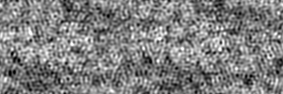
| Projections & slices | Image control
Images are generated by Spider. generated in cubic-lattice coordinate | ||||||||||||||||||||||||||||||||||||||||||||||||||||||||||||||||||||
| Voxel size | X: 1.33333 Å / Y: 1.32143 Å / Z: 1.32951 Å | ||||||||||||||||||||||||||||||||||||||||||||||||||||||||||||||||||||
| Density |
| ||||||||||||||||||||||||||||||||||||||||||||||||||||||||||||||||||||
| Symmetry | Space group: 1 | ||||||||||||||||||||||||||||||||||||||||||||||||||||||||||||||||||||
| Details | EMDB XML:
CCP4 map header:
| ||||||||||||||||||||||||||||||||||||||||||||||||||||||||||||||||||||
 Y (Sec.)
 X (Row.)
 Z (Col.)


-Supplemental data
- Sample components
-Entire : Acrosomal bundle from Limulus sperm
| Entire | Name: Acrosomal bundle from Limulus sperm |
|---|---|
| Components |
|
-Supramolecule #1000: Acrosomal bundle from Limulus sperm
| Supramolecule | Name: Acrosomal bundle from Limulus sperm / type: sample / ID: 1000 Oligomeric state: helix of actin and scruin in a 1 to 1 stoichiometry Number unique components: 2 |
|---|---|
| Molecular weight | Experimental: 4.2 MDa / Theoretical: 4.2 MDa / Method: calculated from molecular weight |
-Macromolecule #1: actin
| Macromolecule | Name: actin / type: protein_or_peptide / ID: 1 / Name.synonym: F-actin / Details: 28 of these per unit cell / Number of copies: 28 / Oligomeric state: helical / Recombinant expression: No / Database: NCBI |
|---|---|
| Source (natural) | Organism: Limulus polyphemus (Atlantic horseshoe crab) / synonym: horseshoe crab / Tissue: sperm / Cell: sperm / Organelle: acrosomal process / Location in cell: acrosomal bundle |
| Molecular weight | Experimental: 1.176 MDa / Theoretical: 1.176 MDa |
-Macromolecule #2: scruin
| Macromolecule | Name: scruin / type: protein_or_peptide / ID: 2 / Name.synonym: scruin / Details: 28 of these per unit cell / Number of copies: 28 / Oligomeric state: helical / Recombinant expression: No / Database: NCBI |
|---|---|
| Source (natural) | Organism: Limulus polyphemus (Atlantic horseshoe crab) / synonym: horseshoe crab / Tissue: sperm / Cell: sperm / Organelle: acrosomal process / Location in cell: acrosomal bundle |
| Molecular weight | Experimental: 3.08 MDa / Theoretical: 3.08 MDa |
-Experimental details
-Structure determination
| Method | cryo EM |
|---|---|
 Processing Processing | helical reconstruction |
| Aggregation state | 2D array |
-Sample preparation
| Concentration | 10 mg/mL |
|---|---|
| Buffer | pH: 7.4 / Details: see J Mol Biol, 221: 711-725 (1991) |
| Vitrification | Cryogen name: ETHANE / Chamber humidity: 100 % / Chamber temperature: 90 K / Instrument: HOMEMADE PLUNGER / Details: Vitrification instrument: homemade guillotine |
| Details | naturally occurring intracellular ordered array. Space group is P2sub1 or P1 21 1. P 1 21 refers to a 2-fold screw axis in the plane of the 2D crystal, whereas I have defined the 2-fold screw along z (second setting monoclinic). |
- Electron microscopy
| Microscope | JEOL 4000EX |
|---|---|
| Temperature | Average: 90 K |
| Image recording | Category: FILM / Film or detector model: KODAK SO-163 FILM / Digitization - Scanner: ZEISS SCAI / Digitization - Sampling interval: 7 µm / Number real images: 90 / Average electron dose: 15 e/Å2 / Details: 153 bundles selected / Od range: 1.5 |
| Electron beam | Acceleration voltage: 400 kV / Electron source: LAB6 |
| Electron optics | Illumination mode: FLOOD BEAM / Imaging mode: BRIGHT FIELD / Nominal defocus max: 3.0 µm / Nominal defocus min: 0.8 µm / Nominal magnification: 40000 |
| Sample stage | Specimen holder: side entry / Specimen holder model: GATAN LIQUID NITROGEN |
-Image processing
| Final reconstruction | Algorithm: OTHER / Resolution.type: BY AUTHOR / Resolution: 9.5 Å / Resolution method: OTHER / Software - Name: MRC, CCP4, homemade Details: 1000 2 unit cell segments from 153 bundles (ref:Schmid, MF, J,. Struct Biol. 2003, 144:195-208. |
|---|---|
| CTF correction | Details: each bundle |
| Crystal parameters | Unit cell - A: 145 Å / Unit cell - B: 145 Å / Unit cell - C: 765 Å / Unit cell - γ: 120 ° / Unit cell - α: 90 ° / Unit cell - β: 90 ° / Plane group: P 1 21 |
-Atomic model buiding 1
| Initial model | PDB ID: |
|---|---|
| Software | Name: foldhunter and homemade software |
| Details | Protocol: cross-correlation. each of the 28 actins was fitted and averaged, scruin was density-correlated and averaged because there is no crystal structure. overall B is the NEGATIVE B factor applied as a high-pass filter in the calculation of the map (-500). |
| Refinement | Space: REAL / Overall B value: -500 / Target criteria: cross-correlation |
| Output model | 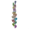 PDB-3b5u:  PDB-3b63: |