ムービー
ムービー コントローラー


+ データを開く

- 基本情報
基本情報
| 登録情報 | データベース: EMDB / ID: EMD-1079 | |||||||||
|---|---|---|---|---|---|---|---|---|---|---|
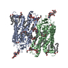
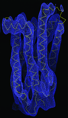
| タイトル | Electron crystallography reveals the structure of metarhodopsin I. | |||||||||
 マップデータ マップデータ | Density map of a rhodopsin photostationary state, highly enriched in metarhodopsin I | |||||||||
 試料 試料 |
| |||||||||
| 機能・相同性 | G protein-coupled opsin signaling pathway => GO:0016056 / Rhodopsin 機能・相同性情報 機能・相同性情報 | |||||||||
| 生物種 |  | |||||||||
| 手法 | 電子線結晶学 / クライオ電子顕微鏡法 / 解像度: 5.5 Å | |||||||||
 データ登録者 データ登録者 | Ruprecht JJ / Mielke T / Vogel R / Villa C / Schertler GFX | |||||||||
 引用 引用 | ジャーナル: EMBO J / 年: 2004 タイトル: Electron crystallography reveals the structure of metarhodopsin I. 著者: Jonathan J Ruprecht / Thorsten Mielke / Reiner Vogel / Claudio Villa / Gebhard F X Schertler /  要旨: Rhodopsin is the prototypical G protein-coupled receptor, responsible for detection of dim light in vision. Upon absorption of a photon, rhodopsin undergoes structural changes, characterised by ...Rhodopsin is the prototypical G protein-coupled receptor, responsible for detection of dim light in vision. Upon absorption of a photon, rhodopsin undergoes structural changes, characterised by distinct photointermediates. Currently, only the ground-state structure has been described. We have determined a density map of a photostationary state highly enriched in metarhodopsin I, to a resolution of 5.5 A in the membrane plane, by electron crystallography. The map shows density for helix 8, the cytoplasmic loops, the extracellular plug, all tryptophan residues, an ordered cholesterol molecule and the beta-ionone ring. Comparison of this map with X-ray structures of the ground state reveals that metarhodopsin I formation does not involve large rigid-body movements of helices, but there is a rearrangement close to the bend of helix 6, at the level of the retinal chromophore. There is no gradual build-up of the large conformational change known to accompany metarhodopsin II formation. The protein remains in a conformation similar to that of the ground state until late in the photobleaching process. | |||||||||
| 履歴 |
|
- 構造の表示
構造の表示
| ムービー |
ムービービューア |
|---|---|
| 構造ビューア | EMマップ: SurfViewMolmilJmol/JSmol |
| 添付画像 |
- ダウンロードとリンク
-EMDBアーカイブ
| マップデータ | emd_1079.map.gz | 14.5 MB | EMDBマップデータ形式 | |
|---|---|---|---|---|
| ヘッダ (付随情報) | emd-1079-v30.xmlemd-1079.xml | 12.3 KB 12.3 KB | 表示 表示 | EMDBヘッダ |
| 画像 |  1079.gif 1079.gif | 73.4 KB | ||
| アーカイブディレクトリ |  http://ftp.pdbj.org/pub/emdb/structures/EMD-1079ftp://ftp.pdbj.org/pub/emdb/structures/EMD-1079 http://ftp.pdbj.org/pub/emdb/structures/EMD-1079ftp://ftp.pdbj.org/pub/emdb/structures/EMD-1079 | HTTPS FTP |
-関連構造データ
| 類似構造データ |
|---|
-リンク
| EMDBのページ | EMDB (EBI/PDBe) / EMDataResource |
|---|
-マップ
| ファイル | ダウンロード / ファイル: emd_1079.map.gz / 形式: CCP4 / 大きさ: 18.5 MB / タイプ: IMAGE STORED AS FLOATING POINT NUMBER (4 BYTES) | ||||||||||||||||||||||||||||||||||||||||||||||||||||||||||||||||||||
|---|---|---|---|---|---|---|---|---|---|---|---|---|---|---|---|---|---|---|---|---|---|---|---|---|---|---|---|---|---|---|---|---|---|---|---|---|---|---|---|---|---|---|---|---|---|---|---|---|---|---|---|---|---|---|---|---|---|---|---|---|---|---|---|---|---|---|---|---|---|
| 注釈 | Density map of a rhodopsin photostationary state, highly enriched in metarhodopsin I | ||||||||||||||||||||||||||||||||||||||||||||||||||||||||||||||||||||
| 投影像・断面図 | 画像のコントロール
画像は Spider により作成 これらの図は立方格子座標系で作成されたものです | ||||||||||||||||||||||||||||||||||||||||||||||||||||||||||||||||||||
| ボクセルのサイズ |
| ||||||||||||||||||||||||||||||||||||||||||||||||||||||||||||||||||||
| 密度 |
| ||||||||||||||||||||||||||||||||||||||||||||||||||||||||||||||||||||
| 対称性 | 空間群: 18 | ||||||||||||||||||||||||||||||||||||||||||||||||||||||||||||||||||||
| 詳細 | EMDB XML:
CCP4マップ ヘッダ情報:
| ||||||||||||||||||||||||||||||||||||||||||||||||||||||||||||||||||||
 Z (Sec.)
 X (Row.)
 Y (Col.)


-添付データ
- 試料の構成要素
-全体 : Bovine Metarhodopsin I
| 全体 | 名称: Bovine Metarhodopsin I |
|---|---|
| 要素 |
|
-超分子 #1000: Bovine Metarhodopsin I
| 超分子 | 名称: Bovine Metarhodopsin I / タイプ: sample / ID: 1000 詳細: Illuminated to form a photostationary state highly enriched in metarhodopsin I 集合状態: monomer / Number unique components: 1 |
|---|---|
| 分子量 | 実験値: 39 KDa / 理論値: 39 KDa |
-分子 #1: Rhodopsin
| 分子 | 名称: Rhodopsin / タイプ: protein_or_peptide / ID: 1 / Name.synonym: Rh / 詳細: Illuminated to form photostationary state / コピー数: 1 / 集合状態: Monomer / 組換発現: No / データベース: NCBI |
|---|---|
| 由来(天然) | 生物種: |
| 分子量 | 実験値: 39 KDa / 理論値: 39 KDa |
| 配列 | GO: G protein-coupled opsin signaling pathway => GO:0016056 / InterPro: Rhodopsin |
-実験情報
-構造解析
| 手法 | クライオ電子顕微鏡法 |
|---|---|
 解析 解析 | 電子線結晶学 |
| 試料の集合状態 | 2D array |
-試料調製
| 濃度 | 0.5 mg/mL |
|---|---|
| 緩衝液 | pH: 7 詳細: 20mM HEPES pH 7, 100 mM NaCl, 10 mM MgCl2, 3 mM NaN3, 4 mM DTT, 4 mM mercaptoethanol, 2.5 % (v/v) isopropanol |
| グリッド | 詳細: 300 mesh molybdenum grid |
| 凍結 | 凍結剤: ETHANE / チャンバー内湿度: 75 % / チャンバー内温度: 77 K / 装置: GATAN CRYOPLUNGE 3 詳細: Vitrification instrument: Gatan cryoplunge. Vitrification carried out in temperature and humidity-controlled chamber Timed resolved state: Vitrified after illumination of specimen 手法: Blot for 20 sec before plunging |
| 詳細 | 11 day dialysis at 18 deg C |
| 結晶化 | 詳細: 11 day dialysis at 18 deg C |
- 電子顕微鏡法
| 顕微鏡 | FEI TECNAI F30 |
|---|---|
| 温度 | 最低: 77 K / 最高: 77 K / 平均: 77 K |
| アライメント法 | Legacy - 非点収差: corrected at 230,000 times magnification |
| 撮影 | カテゴリ: FILM / フィルム・検出器のモデル: KODAK SO-163 FILM / デジタル化 - スキャナー: ZEISS SCAI / デジタル化 - サンプリング間隔: 7 µm / 実像数: 87 / 平均電子線量: 15 e/Å2 |
| Tilt angle min | 0 |
| 電子線 | 加速電圧: 300 kV / 電子線源:  FIELD EMISSION GUN FIELD EMISSION GUN |
| 電子光学系 | 倍率(補正後): 59000 / 照射モード: SPOT SCAN / 撮影モード: BRIGHT FIELD / Cs: 2.3 mm / 最大 デフォーカス(公称値): 1.41 µm / 最小 デフォーカス(公称値): 0.27 µm / 倍率(公称値): 60000 |
| 試料ステージ | 試料ホルダー: Side entry liquid-nitrogen cooled Gatan holder 試料ホルダーモデル: GATAN LIQUID NITROGEN / Tilt angle max: 60 / Tilt series - Axis1 - Min angle: 0 ° / Tilt series - Axis1 - Max angle: 60 ° |
| 実験機器 |  モデル: Tecnai F30 / 画像提供: FEI Company |
-画像解析
| 最終 再構成 | アルゴリズム: OTHER / 解像度のタイプ: BY AUTHOR / 解像度: 5.5 Å / 解像度の算出法: OTHER / ソフトウェア - 名称: MRC |
|---|---|
| 結晶パラメータ | 単位格子 - A: 58.8 Å / 単位格子 - B: 83.7 Å / 単位格子 - C: 200.0 Å / 単位格子 - γ: 90 ° / 単位格子 - α: 90 ° / 単位格子 - β: 90 ° / 面群: P 2 21 21 |
| CTF補正 | 詳細: Each image |
-原子モデル構築 1
| 初期モデル | PDB ID: Chain - Chain ID: B |
|---|---|
| ソフトウェア | 名称: O |
| 詳細 | PDBEntryID_givenInChain. Protocol: Rigid Body |
| 精密化 | 空間: REAL / プロトコル: RIGID BODY FIT |