Movie
Movie Controller

[English] 日本語
 Yorodumi
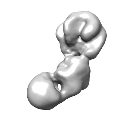
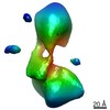
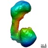
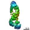
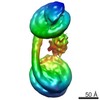
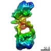
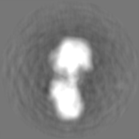
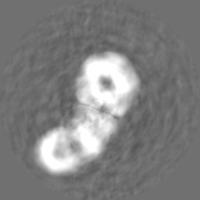
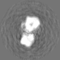
- EMDB-10630: Interaction of the CoREST complex with a nucleosome with 185 bp 6... -
+ Open data

- Basic information
| Entry | Database: EMDB / ID: EMD-10630 | |||||||||
|---|---|---|---|---|---|---|---|---|---|---|
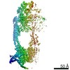
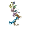
| Title | Interaction of the CoREST complex with a nucleosome with 185 bp 601 sequence DNA and a propargylamine mimic of dimethy Lys4 histone H3 | |||||||||
 Map data Map data | ||||||||||
 Sample Sample |
| |||||||||
 Keywords Keywords | CoREST / RCOR1 / HDAC1 / LSD1 / nucleosome / GENE REGULATION | |||||||||
| Biological species |  Homo sapiens (human) Homo sapiens (human) | |||||||||
| Method | single particle reconstruction / negative staining / Resolution: 26.1 Å | |||||||||
 Authors Authors | Song Y / Fairall L / Wu M / Ragan TJ / Savva CG / Morone N / Cole PA / Schwabe JWR | |||||||||
| Funding support |  United Kingdom, 1 items United Kingdom, 1 items
| |||||||||
 Citation Citation | Journal: Cell Rep / Year: 2020 Title: Mechanism of Crosstalk between the LSD1 Demethylase and HDAC1 Deacetylase in the CoREST Complex. Authors: Yun Song / Lisbeth Dagil / Louise Fairall / Naomi Robertson / Mingxuan Wu / T J Ragan / Christos G Savva / Almutasem Saleh / Nobuhiro Morone / Micha B A Kunze / Andrew G Jamieson / Philip A ...Authors: Yun Song / Lisbeth Dagil / Louise Fairall / Naomi Robertson / Mingxuan Wu / T J Ragan / Christos G Savva / Almutasem Saleh / Nobuhiro Morone / Micha B A Kunze / Andrew G Jamieson / Philip A Cole / D Flemming Hansen / John W R Schwabe /  Abstract: The transcriptional corepressor complex CoREST is one of seven histone deacetylase complexes that regulate the genome through controlling chromatin acetylation. The CoREST complex is unique in ...The transcriptional corepressor complex CoREST is one of seven histone deacetylase complexes that regulate the genome through controlling chromatin acetylation. The CoREST complex is unique in containing both histone demethylase and deacetylase enzymes, LSD1 and HDAC1, held together by the RCOR1 scaffold protein. To date, it has been assumed that the enzymes function independently within the complex. Now, we report the assembly of the ternary complex. Using both structural and functional studies, we show that the activity of the two enzymes is closely coupled and that the complex can exist in at least two distinct states with different kinetics. Electron microscopy of the complex reveals a bi-lobed structure with LSD1 and HDAC1 enzymes at opposite ends of the complex. The structure of CoREST in complex with a nucleosome reveals a mode of chromatin engagement that contrasts with previous models. | |||||||||
| History |
|
- Structure visualization
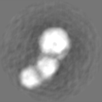
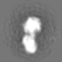
| Movie |
 Movie viewer Movie viewer |
|---|---|
| Structure viewer | EM map: SurfViewMolmilJmol/JSmol |
| Supplemental images |
- Downloads & links
-EMDB archive
| Map data | emd_10630.map.gz | 23.3 MB | EMDB map data format | |
|---|---|---|---|---|
| Header (meta data) | emd-10630-v30.xmlemd-10630.xml | 16.9 KB 16.9 KB | Display Display | EMDB header |
| Images |  emd_10630.png emd_10630.png | 35.3 KB | ||
| Filedesc metadata | emd-10630.cif.gz | 4.6 KB | ||
| Others | emd_10630_half_map_1.map.gzemd_10630_half_map_2.map.gz | 23.4 MB 23.4 MB | ||
| Archive directory |  http://ftp.pdbj.org/pub/emdb/structures/EMD-10630ftp://ftp.pdbj.org/pub/emdb/structures/EMD-10630 http://ftp.pdbj.org/pub/emdb/structures/EMD-10630ftp://ftp.pdbj.org/pub/emdb/structures/EMD-10630 | HTTPS FTP |
-Related structure data
| Related structure data | C: citing same article ( |
|---|---|
| Similar structure data |


-Links
| EMDB pages | EMDB (EBI/PDBe) / EMDataResource |
|---|
-Map
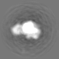
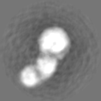
| File | Download / File: emd_10630.map.gz / Format: CCP4 / Size: 30.5 MB / Type: IMAGE STORED AS FLOATING POINT NUMBER (4 BYTES) | ||||||||||||||||||||||||||||||||||||||||||||||||||||||||||||
|---|---|---|---|---|---|---|---|---|---|---|---|---|---|---|---|---|---|---|---|---|---|---|---|---|---|---|---|---|---|---|---|---|---|---|---|---|---|---|---|---|---|---|---|---|---|---|---|---|---|---|---|---|---|---|---|---|---|---|---|---|---|
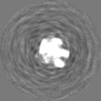
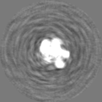
| Projections & slices | Image control
Images are generated by Spider. | ||||||||||||||||||||||||||||||||||||||||||||||||||||||||||||
| Voxel size | X=Y=Z: 1.83 Å | ||||||||||||||||||||||||||||||||||||||||||||||||||||||||||||
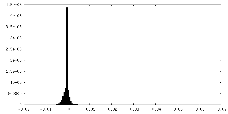
| Density |
| ||||||||||||||||||||||||||||||||||||||||||||||||||||||||||||
| Symmetry | Space group: 1 | ||||||||||||||||||||||||||||||||||||||||||||||||||||||||||||
| Details | EMDB XML:
CCP4 map header:
| ||||||||||||||||||||||||||||||||||||||||||||||||||||||||||||
 Z (Sec.)
 Y (Row.)
 X (Col.)


-Supplemental data
-Half map: #1
| File | emd_10630_half_map_1.map | ||||||||||||
|---|---|---|---|---|---|---|---|---|---|---|---|---|---|
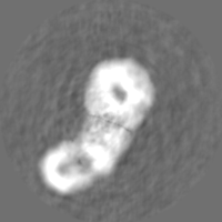
| Projections & Slices |
| ||||||||||||
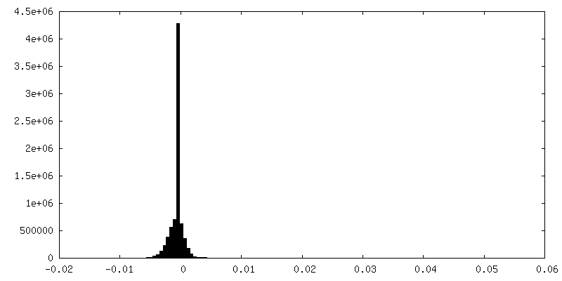
| Density Histograms |
-Half map: #2
| File | emd_10630_half_map_2.map | ||||||||||||
|---|---|---|---|---|---|---|---|---|---|---|---|---|---|
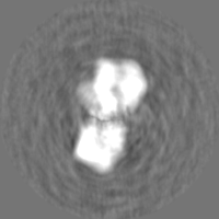
| Projections & Slices |
| ||||||||||||
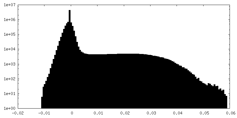
| Density Histograms |

- Sample components
-Entire : Nucleosome with CoREST complex (RCOR1: LSD1:HDAC1)
| Entire | Name: Nucleosome with CoREST complex (RCOR1: LSD1:HDAC1) |
|---|---|
| Components |
|
-Supramolecule #1: Nucleosome with CoREST complex (RCOR1: LSD1:HDAC1)
| Supramolecule | Name: Nucleosome with CoREST complex (RCOR1: LSD1:HDAC1) / type: complex / ID: 1 / Parent: 0 |
|---|---|
| Source (natural) | Organism: Homo sapiens (human) |
| Molecular weight | Theoretical: 410 KDa |
-Experimental details
-Structure determination
| Method | negative staining |
|---|---|
 Processing Processing | single particle reconstruction |
| Aggregation state | particle |
-Sample preparation
| Concentration | 0.03 mg/mL | |||||||||
|---|---|---|---|---|---|---|---|---|---|---|
| Buffer | pH: 7.5 Component:
| |||||||||
| Staining | Type: NEGATIVE / Material: uranyl acetate | |||||||||
| Grid | Model: C-flat / Material: COPPER / Mesh: 400 / Support film - Material: CARBON / Support film - topology: CONTINUOUS / Pretreatment - Type: GLOW DISCHARGE | |||||||||
| Details | Sample was crosslinked with 0.01% glutaraldehyde |
- Electron microscopy
| Microscope | TFS TALOS |
|---|---|
| Image recording | Film or detector model: FEI CETA (4k x 4k) / Detector mode: COUNTING / Digitization - Dimensions - Width: 4096 pixel / Digitization - Dimensions - Height: 4096 pixel / Digitization - Frames/image: 1-48 / Number grids imaged: 1 / Number real images: 384 / Average exposure time: 1.0 sec. / Average electron dose: 35.0 e/Å2 |
| Electron beam | Acceleration voltage: 200 kV / Electron source:  FIELD EMISSION GUN FIELD EMISSION GUN |
| Electron optics | Illumination mode: FLOOD BEAM / Imaging mode: BRIGHT FIELD / Cs: 2.7 mm / Nominal defocus max: 2.0 µm / Nominal defocus min: 1.0 µm |
| Sample stage | Specimen holder model: SIDE ENTRY, EUCENTRIC |