 Movie
Movie Controller

[English] 日本語
 Yorodumi
- EMDB-1060: Electron cryomicroscopy and bioinformatics suggest protein fold m... -
+ Open data

- Basic information
| Entry | Database: EMDB / ID: EMD-1060 | |||||||||
|---|---|---|---|---|---|---|---|---|---|---|
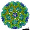
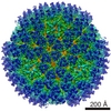
| Title | Electron cryomicroscopy and bioinformatics suggest protein fold models for rice dwarf virus. | |||||||||
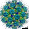
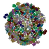
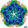
 Map data Map data | This is the 3f average map from Rice Dwarf Virus | |||||||||
 Sample Sample |
| |||||||||
| Biological species |  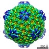 Rice dwarf virus Rice dwarf virus | |||||||||
| Method | single particle reconstruction / cryo EM / Resolution: 6.8 Å | |||||||||
 Authors Authors | Zhou ZH / Baker ML / Jiang W / Dougherty M / Jakana J / Dong G / Lu G / Chiu W | |||||||||
 Citation Citation | Journal: Nat Struct Biol / Year: 2001 Title: Electron cryomicroscopy and bioinformatics suggest protein fold models for rice dwarf virus. Authors: Z H Zhou / M L Baker / W Jiang / M Dougherty / J Jakana / G Dong / G Lu / W Chiu /  Abstract: The three-dimensional structure of rice dwarf virus was determined to 6.8 A resolution by single particle electron cryomicroscopy. By integrating the structural analysis with bioinformatics, the ...The three-dimensional structure of rice dwarf virus was determined to 6.8 A resolution by single particle electron cryomicroscopy. By integrating the structural analysis with bioinformatics, the folds of the proteins in the double-shelled capsid were derived. In the outer shell protein, the uniquely orientated upper and lower domains are composed of similar secondary structure elements but have different relative orientations from that of bluetongue virus in the same Reoviridae family. Differences in both sequence and structure between these proteins may be important in defining virus-host interactions. The inner shell protein adopts a conformation similar to other members of Reoviridae, suggesting a common ancestor that has evolved to infect hosts ranging from plants to animals. Symmetry mismatch between the two shells results in nonequivalent, yet specific, interactions that contribute to the stability of this large macromolecular machine. | |||||||||
| History |
|
- Structure visualization
| Movie |
 Movie viewer Movie viewer |
|---|---|
| Structure viewer | EM map: SurfViewMolmilJmol/JSmol |
| Supplemental images |
 UCSF Chimera
- Downloads & links
-EMDB archive
| Map data | emd_1060.map.gz | 21.6 MB | EMDB map data format | |
|---|---|---|---|---|
| Header (meta data) | emd-1060-v30.xmlemd-1060.xml | 9.5 KB 9.5 KB | Display Display | EMDB header |
| Images |  1060.gif 1060.gif | 93.1 KB | ||
| Others | emd_1060_additional_1.map.gzemd_1060_additional_2.map.gz | 835.9 KB 130.9 KB | ||
| Archive directory |  http://ftp.pdbj.org/pub/emdb/structures/EMD-1060ftp://ftp.pdbj.org/pub/emdb/structures/EMD-1060 http://ftp.pdbj.org/pub/emdb/structures/EMD-1060ftp://ftp.pdbj.org/pub/emdb/structures/EMD-1060 | HTTPS FTP |
-Related structure data
| Similar structure data |
|---|

-Links
| EMDB pages | EMDB (EBI/PDBe) / EMDataResource |
|---|
-Map
| File | Download / File: emd_1060.map.gz / Format: CCP4 / Size: 40.6 MB / Type: IMAGE STORED AS FLOATING POINT NUMBER (4 BYTES) | ||||||||||||||||||||||||||||||||||||||||||||||||||||||||||||||||||||
|---|---|---|---|---|---|---|---|---|---|---|---|---|---|---|---|---|---|---|---|---|---|---|---|---|---|---|---|---|---|---|---|---|---|---|---|---|---|---|---|---|---|---|---|---|---|---|---|---|---|---|---|---|---|---|---|---|---|---|---|---|---|---|---|---|---|---|---|---|---|
| Annotation | This is the 3f average map from Rice Dwarf Virus | ||||||||||||||||||||||||||||||||||||||||||||||||||||||||||||||||||||
| Projections & slices | Image control
Images are generated by Spider. generated in cubic-lattice coordinate | ||||||||||||||||||||||||||||||||||||||||||||||||||||||||||||||||||||
| Voxel size | X=Y=Z: 1.64 Å | ||||||||||||||||||||||||||||||||||||||||||||||||||||||||||||||||||||
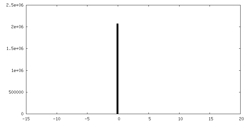
| Density |
| ||||||||||||||||||||||||||||||||||||||||||||||||||||||||||||||||||||
| Symmetry | Space group: 1 | ||||||||||||||||||||||||||||||||||||||||||||||||||||||||||||||||||||
| Details | EMDB XML:
CCP4 map header:
| ||||||||||||||||||||||||||||||||||||||||||||||||||||||||||||||||||||
 Z (Sec.)
 Y (Row.)
 X (Col.)


-Supplemental data
-Supplemental map: emd 1060 additional 1.map
| File | emd_1060_additional_1.map | ||||||||||||
|---|---|---|---|---|---|---|---|---|---|---|---|---|---|
| Projections & Slices |
| ||||||||||||
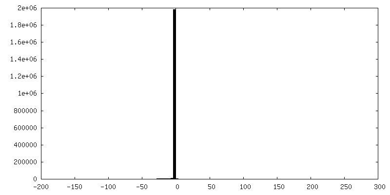
| Density Histograms |


-Supplemental map: emd 1060 additional 2.map
| File | emd_1060_additional_2.map | ||||||||||||
|---|---|---|---|---|---|---|---|---|---|---|---|---|---|
| Projections & Slices |
| ||||||||||||
| Density Histograms |


-Supplemental map: emd 1060 averaged monomer.mrc
| File | emd_1060_averaged_monomer.mrc | ||||||||||||
|---|---|---|---|---|---|---|---|---|---|---|---|---|---|
| Projections & Slices |
| ||||||||||||
| Density Histograms |


-Supplemental map: emd 1060 averaged trimer.mrc
| File | emd_1060_averaged_trimer.mrc | ||||||||||||
|---|---|---|---|---|---|---|---|---|---|---|---|---|---|
| Projections & Slices |
| ||||||||||||
| Density Histograms |


- Sample components
-Entire : Rice Dwarf Virus
| Entire | Name: Rice Dwarf Virus |
|---|---|
| Components |
|
-Supramolecule #1000: Rice Dwarf Virus
| Supramolecule | Name: Rice Dwarf Virus / type: sample / ID: 1000 / Details: Full capsid / Number unique components: 1 |
|---|---|
| Molecular weight | Theoretical: 50 MDa |
-Supramolecule #1: Rice dwarf virus
| Supramolecule | Name: Rice dwarf virus / type: virus / ID: 1 / Name.synonym: rdv / NCBI-ID: 10991 / Sci species name: Rice dwarf virus / Virus type: VIRION / Virus isolate: STRAIN / Virus enveloped: No / Virus empty: No / Syn species name: rdv |
|---|---|
| Host (natural) | synonym: PLANTAE(HIGHER PLANTS) |
| Virus shell | Shell ID: 1 / Name: P3 / Diameter: 590 Å / T number (triangulation number): 1 |
| Virus shell | Shell ID: 2 / Name: P8 / Diameter: 700 Å / T number (triangulation number): 13 |
-Experimental details
-Structure determination
| Method | cryo EM |
|---|---|
 Processing Processing | single particle reconstruction |
| Aggregation state | particle |
-Sample preparation
| Vitrification | Cryogen name: ETHANE / Chamber humidity: 27 % / Chamber temperature: 111 K / Instrument: HOMEMADE PLUNGER / Details: Vitrification instrument: manual |
|---|
- Electron microscopy
| Microscope | JEOL 4000 |
|---|---|
| Temperature | Average: 109 K |
| Details | Microscope:JEOL 4000 |
| Date | Jan 1, 1999 |
| Image recording | Category: FILM / Film or detector model: KODAK SO-163 FILM / Digitization - Scanner: ZEISS SCAI / Digitization - Sampling interval: 14 µm / Number real images: 162 / Average electron dose: 13 e/Å2 / Camera length: 200 / Bits/pixel: 12 |
| Electron beam | Acceleration voltage: 400 kV / Electron source: LAB6 |
| Electron optics | Calibrated magnification: 49495 / Illumination mode: FLOOD BEAM / Imaging mode: BRIGHT FIELD / Cs: 4.1 mm / Nominal defocus max: 2.2 µm / Nominal defocus min: 0.3 µm / Nominal magnification: 50000 |
| Sample stage | Specimen holder: Gatan / Specimen holder model: GATAN LIQUID NITROGEN |
-Image processing
| CTF correction | Details: per micrograph |
|---|---|
| Final reconstruction | Applied symmetry - Point group: I (icosahedral) / Algorithm: OTHER / Resolution.type: BY AUTHOR / Resolution: 6.8 Å / Resolution method: FSC 0.5 CUT-OFF / Software - Name: MRC,IMRS / Number images used: 3261 |
-Atomic model buiding 1
| Initial model | PDB ID: |
|---|---|
| Software | Name: Foldhunter |
| Details | FOLDHUNTER is decribed in Jiang et al , J.Mol.Biol., 308, 1033-1044 (2001). Beta sheet domain BTV VP7 blurred to 7 angstroms using the program PDB2MRC (Ludtke et at, J.Struct.Biol.,128, 82-97 (1999). |
| Refinement | Overall B value: 100 |

