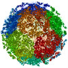
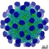
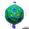
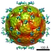
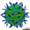
ジャーナル: J Biol Chem / 年: 2004 タイトル: The structure of echovirus type 12 bound to a two-domain fragment of its cellular attachment protein decay-accelerating factor (CD 55). 著者: David Bhella / Ian G Goodfellow / Pietro Roversi / David Pettigrew / Yasmin Chaudhry / David J Evans / Susan M Lea / 要旨: Echovirus type 12 (EV12), an Enterovirus of the Picornaviridae family, uses the complement regulator decay-accelerating factor (DAF, CD55) as a cellular receptor. We have calculated a three- ...Echovirus type 12 (EV12), an Enterovirus of the Picornaviridae family, uses the complement regulator decay-accelerating factor (DAF, CD55) as a cellular receptor. We have calculated a three-dimensional reconstruction of EV12 bound to a fragment of DAF consisting of short consensus repeat domains 3 and 4 from cryo-negative stain electron microscopy data (EMD code 1057). This shows that, as for an earlier reconstruction of the related echovirus type 7 bound to DAF, attachment is not within the viral canyon but occurs close to the 2-fold symmetry axes. Despite this general similarity our reconstruction reveals a receptor interaction that is quite different from that observed for EV7. Fitting of the crystallographic co-ordinates for DAF(34) and EV11 into the reconstruction shows a close agreement between the crystal structure of the receptor fragment and the density for the virus-bound receptor, allowing unambiguous positioning of the receptor with respect to the virion (PDB code 1UPN). Our finding that the mode of virus-receptor interaction in EV12 is distinct from that seen for EV7 raises interesting questions regarding the evolution and biological significance of the DAF binding phenotype in these viruses.
詳細: merge of focal pair images of single particles
最終 再構成
想定した対称性 - 点群: I (正20面体型対称) / アルゴリズム: OTHER / 解像度のタイプ: BY AUTHOR / 解像度: 18.0 Å / 解像度の算出法: FSC 0.5 CUT-OFF / ソフトウェア - 名称: EM3DR2, PFT2, CTFMIX 詳細: Particles were aligned using a model based strategy starting with a model derived from the crystallographic co-ordinates of EV-1, filtered to 16 Angstroms resolution. The program is called ...詳細: Particles were aligned using a model based strategy starting with a model derived from the crystallographic co-ordinates of EV-1, filtered to 16 Angstroms resolution. The program is called PFT (Polar Fourier Transform). The reconstructions were calculated using the EM3DR2 program which is based on the standard method of calculating icosahedral reconstructions as described by Crowther,'Fourier-Bessel'. 使用した粒子像数: 617
-
原子モデル構築 1
詳細
3D crystal structure fitting details lodged with PDB 1UPN
得られたモデル
PDB-1upn: COMPLEX OF ECHOVIRUS TYPE 12 WITH DOMAINS 3 AND 4 OF ITS RECEPTOR DECAY ACCELERATING FACTOR (CD55) BY CRYO ELECTRON MICROSCOPY AT 16 A
 ムービー
ムービー コントローラー


 データを開く
 基本情報
基本情報 マップデータ
 試料
試料 機能・相同性情報
 Human echovirus 12 (ウイルス)
 データ登録者
 引用
引用
 構造の表示
構造の表示 UCSF Chimera
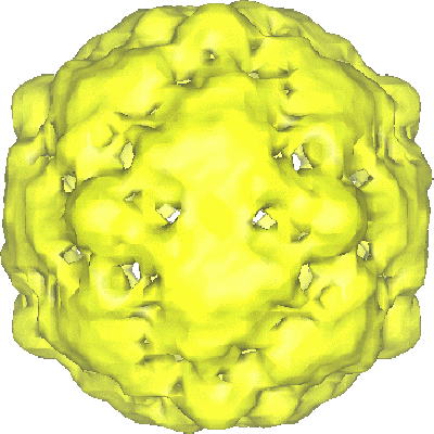
 ダウンロードとリンク
 1058.gif
 http://ftp.pdbj.org/pub/emdb/structures/EMD-1058
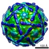


 Z (Sec.)
 Y (Row.)
 X (Col.)


 試料の構成要素
 Homo sapiens (ヒト) / 別称: VERTEBRATES
 解析
解析 電子顕微鏡法