 ムービー
ムービー コントローラー


+ データを開く

- 基本情報
基本情報
| 登録情報 | データベース: EMDB / ID: EMD-1050 | |||||||||
|---|---|---|---|---|---|---|---|---|---|---|
| タイトル | The cyanide degrading nitrilase from Pseudomonas stutzeri AK61 is a two-fold symmetric, 14-subunit spiral. | |||||||||
 マップデータ マップデータ | ||||||||||
 試料 試料 |
| |||||||||
| 機能・相同性 | Nitrilase/cyanide hydratase, conserved site / nitrilase activity 機能・相同性情報 機能・相同性情報 | |||||||||
| 生物種 |  Pseudomonas stutzeri (バクテリア) Pseudomonas stutzeri (バクテリア) | |||||||||
| 手法 | 単粒子再構成法 / ネガティブ染色法 / 解像度: 25.0 Å | |||||||||
 データ登録者 データ登録者 | Sewell BT / Berman MN / Meyers PR / Jandhyala D / Benedik MJ | |||||||||
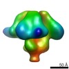
 引用 引用 | ジャーナル: Structure / 年: 2003 タイトル: The cyanide degrading nitrilase from Pseudomonas stutzeri AK61 is a two-fold symmetric, 14-subunit spiral. 著者: B T Sewell / M N Berman / P R Meyers / D Jandhyala / M J Benedik /  要旨: The quaternary structure of the cyanide dihydratase from Pseudomonas stutzeri AK61 was determined by negative stain electron microscopy and three-dimensional reconstruction using the single particle ...The quaternary structure of the cyanide dihydratase from Pseudomonas stutzeri AK61 was determined by negative stain electron microscopy and three-dimensional reconstruction using the single particle technique. The structure is a spiral comprising 14 subunits with 2-fold symmetry. Interactions across the groove cause a decrease in the radius of the spiral at the ends and the resulting steric hindrance prevents the addition of further subunits. Similarity to two members of the nitrilase superfamily, the Nit domain of NitFhit and N-carbamyl-D-amino acid amidohydrolase, enabled the construction of a partial atomic model that could be unambiguously fitted to the stain envelope. The model suggests that interactions involving two significant insertions in the sequence relative to these structures leads to the left-handed spiral assembly. | |||||||||
| 履歴 |
|
- 構造の表示
構造の表示
| ムービー |
ムービービューア |
|---|---|
| 構造ビューア | EMマップ: SurfViewMolmilJmol/JSmol |
| 添付画像 |
 UCSF Chimera
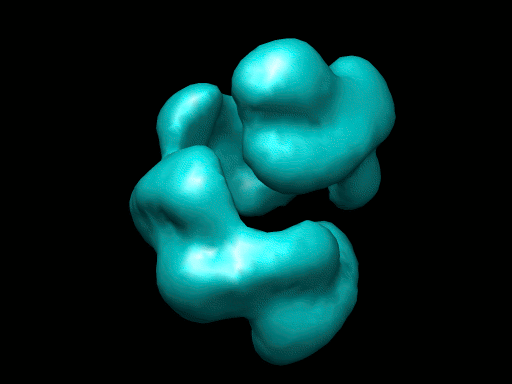
- ダウンロードとリンク
-EMDBアーカイブ
| マップデータ | emd_1050.map.gz | 863.1 KB | EMDBマップデータ形式 | |
|---|---|---|---|---|
| ヘッダ (付随情報) | emd-1050-v30.xmlemd-1050.xml | 11.2 KB 11.2 KB | 表示 表示 | EMDBヘッダ |
| 画像 |  1050.gif 1050.gif | 45.7 KB | ||
| アーカイブディレクトリ |  http://ftp.pdbj.org/pub/emdb/structures/EMD-1050ftp://ftp.pdbj.org/pub/emdb/structures/EMD-1050 http://ftp.pdbj.org/pub/emdb/structures/EMD-1050ftp://ftp.pdbj.org/pub/emdb/structures/EMD-1050 | HTTPS FTP |
-関連構造データ


-リンク
| EMDBのページ | EMDB (EBI/PDBe) / EMDataResource |
|---|
-マップ
| ファイル | ダウンロード / ファイル: emd_1050.map.gz / 形式: CCP4 / 大きさ: 1.9 MB / タイプ: IMAGE STORED AS FLOATING POINT NUMBER (4 BYTES) | ||||||||||||||||||||||||||||||||||||||||||||||||||||||||||||||||||||
|---|---|---|---|---|---|---|---|---|---|---|---|---|---|---|---|---|---|---|---|---|---|---|---|---|---|---|---|---|---|---|---|---|---|---|---|---|---|---|---|---|---|---|---|---|---|---|---|---|---|---|---|---|---|---|---|---|---|---|---|---|---|---|---|---|---|---|---|---|---|
| 投影像・断面図 | 画像のコントロール
画像は Spider により作成 | ||||||||||||||||||||||||||||||||||||||||||||||||||||||||||||||||||||
| ボクセルのサイズ | X=Y=Z: 4 Å | ||||||||||||||||||||||||||||||||||||||||||||||||||||||||||||||||||||
| 密度 |
| ||||||||||||||||||||||||||||||||||||||||||||||||||||||||||||||||||||
| 対称性 | 空間群: 1 | ||||||||||||||||||||||||||||||||||||||||||||||||||||||||||||||||||||
| 詳細 | EMDB XML:
CCP4マップ ヘッダ情報:
| ||||||||||||||||||||||||||||||||||||||||||||||||||||||||||||||||||||
 Z (Sec.)
 Y (Row.)
 X (Col.)


-添付データ
- 試料の構成要素
-全体 : Cyanide dihydratase from Pseudomonas stutzeri
| 全体 | 名称: Cyanide dihydratase from Pseudomonas stutzeri |
|---|---|
| 要素 |
|
-超分子 #1000: Cyanide dihydratase from Pseudomonas stutzeri
| 超分子 | 名称: Cyanide dihydratase from Pseudomonas stutzeri / タイプ: sample / ID: 1000 詳細: The sample eluted as a single symmetric peak on gel filtration chromatography. Three bands were seen on SDS-PAGE presumably indicating some proteolysis 集合状態: 14 identical subunits / Number unique components: 1 |
|---|---|
| 分子量 | 実験値: 500 KDa / 理論値: 532 KDa 手法: gel filtration chromatography, sequencing and counting subunits. |
-分子 #1: cyanide dihydratase
| 分子 | 名称: cyanide dihydratase / タイプ: protein_or_peptide / ID: 1 / Name.synonym: nitrilase 詳細: The protein elutes as a sharp symmetric peak on Sephacryl 300 HR chromatography and particles appear homogeneously sized by negative stain EM コピー数: 14 / 集合状態: 14mer / 組換発現: Yes |
|---|---|
| 由来(天然) | 生物種: Pseudomonas stutzeri (バクテリア) / 株: AK61 / 細胞: bacteria / Organelle: bacteria / 細胞中の位置: cytoplasm |
| 分子量 | 実験値: 38 KDa / 理論値: 38 KDa |
| 組換発現 | 生物種: |
| 配列 | GO: nitrilase activity / InterPro: Nitrilase/cyanide hydratase, conserved site |
-実験情報
-構造解析
| 手法 | ネガティブ染色法 |
|---|---|
 解析 解析 | 単粒子再構成法 |
| 試料の集合状態 | particle |
-試料調製
| 濃度 | 0.3 mg/mL |
|---|---|
| 緩衝液 | pH: 8 / 詳細: 10 mM triethanolamine, 50 mM NaCl |
| 染色 | タイプ: NEGATIVE 詳細: A carbon-coated copper grid was placed on a droplet of this enzyme preparation for 10 minutes, blotted, and then stained with 2% uranyl acetate for 3 minutes. The grid was then blotted and air-dried. |
| グリッド | 詳細: carbon film on 200 mesh copper grid |
| 凍結 | 凍結剤: NONE |
- 電子顕微鏡法
| 顕微鏡 | JEOL 2000EXII |
|---|---|
| 詳細 | Additional details about microscope model:JEOL 1200EXII |
| 日付 | 2001年3月1日 |
| 撮影 | カテゴリ: CCD / フィルム・検出器のモデル: KODAK SO-163 FILM / デジタル化 - サンプリング間隔: 20 µm / 実像数: 15 / ビット/ピクセル: 16 |
| 電子線 | 加速電圧: 120 kV / 電子線源: TUNGSTEN HAIRPIN |
| 電子光学系 | 照射モード: FLOOD BEAM / 撮影モード: BRIGHT FIELD / Cs: 5.6 mm / 最大 デフォーカス(公称値): 0.7 µm / 最小 デフォーカス(公称値): 0.1 µm / 倍率(公称値): 50000 |
| 試料ステージ | 試料ホルダー: Eucentric / 試料ホルダーモデル: OTHER |
+画像解析
-原子モデル構築 1
| ソフトウェア | 名称: CoLoRes, SITUS |
|---|---|
| 詳細 | The map was fitted using a model generated from the Nit structure (1ems in PDB). Magification was refined by dtermining the CoLoRes correlation coefficient as a function of scale factor. Locations of the 14 subunits were verified by vizualization with O. Fitted co-ordinates are available on request.Initially no symmetry was assumed but a two fold axis became apparent after 47 cycles. The orientation of the twofold axis was determined and the model was rotated so that the twofold axis was parallel to the x-axis. In 17 subsequent cycles of refinement the twofold symmetry was imposed by rotating about the x-axis and adding the structure thus formed to the original. |
| 精密化 | プロトコル: RIGID BODY FIT |
