 Movie
Movie Controller

[English] 日本語
 Yorodumi
- EMDB-1050: The cyanide degrading nitrilase from Pseudomonas stutzeri AK61 is... -
+ Open data

- Basic information
| Entry | Database: EMDB / ID: EMD-1050 | |||||||||
|---|---|---|---|---|---|---|---|---|---|---|
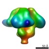
| Title | The cyanide degrading nitrilase from Pseudomonas stutzeri AK61 is a two-fold symmetric, 14-subunit spiral. | |||||||||
 Map data Map data | ||||||||||
 Sample Sample |
| |||||||||
| Function / homology | Nitrilase/cyanide hydratase, conserved site / nitrilase activity Function and homology information Function and homology information | |||||||||
| Biological species |  Pseudomonas stutzeri (bacteria) Pseudomonas stutzeri (bacteria) | |||||||||
| Method | single particle reconstruction / negative staining / Resolution: 25.0 Å | |||||||||
 Authors Authors | Sewell BT / Berman MN / Meyers PR / Jandhyala D / Benedik MJ | |||||||||
 Citation Citation | Journal: Structure / Year: 2003 Title: The cyanide degrading nitrilase from Pseudomonas stutzeri AK61 is a two-fold symmetric, 14-subunit spiral. Authors: B T Sewell / M N Berman / P R Meyers / D Jandhyala / M J Benedik /  Abstract: The quaternary structure of the cyanide dihydratase from Pseudomonas stutzeri AK61 was determined by negative stain electron microscopy and three-dimensional reconstruction using the single particle ...The quaternary structure of the cyanide dihydratase from Pseudomonas stutzeri AK61 was determined by negative stain electron microscopy and three-dimensional reconstruction using the single particle technique. The structure is a spiral comprising 14 subunits with 2-fold symmetry. Interactions across the groove cause a decrease in the radius of the spiral at the ends and the resulting steric hindrance prevents the addition of further subunits. Similarity to two members of the nitrilase superfamily, the Nit domain of NitFhit and N-carbamyl-D-amino acid amidohydrolase, enabled the construction of a partial atomic model that could be unambiguously fitted to the stain envelope. The model suggests that interactions involving two significant insertions in the sequence relative to these structures leads to the left-handed spiral assembly. | |||||||||
| History |
|
- Structure visualization
| Movie |
Movie viewer |
|---|---|
| Structure viewer | EM map: SurfViewMolmilJmol/JSmol |
| Supplemental images |
 UCSF Chimera
- Downloads & links
-EMDB archive
| Map data | emd_1050.map.gz | 863.1 KB | EMDB map data format | |
|---|---|---|---|---|
| Header (meta data) | emd-1050-v30.xmlemd-1050.xml | 11.2 KB 11.2 KB | Display Display | EMDB header |
| Images |  1050.gif 1050.gif | 45.7 KB | ||
| Archive directory |  http://ftp.pdbj.org/pub/emdb/structures/EMD-1050ftp://ftp.pdbj.org/pub/emdb/structures/EMD-1050 http://ftp.pdbj.org/pub/emdb/structures/EMD-1050ftp://ftp.pdbj.org/pub/emdb/structures/EMD-1050 | HTTPS FTP |
-Related structure data
| Similar structure data |
|---|


-Links
| EMDB pages | EMDB (EBI/PDBe) / EMDataResource |
|---|
-Map
| File | Download / File: emd_1050.map.gz / Format: CCP4 / Size: 1.9 MB / Type: IMAGE STORED AS FLOATING POINT NUMBER (4 BYTES) | ||||||||||||||||||||||||||||||||||||||||||||||||||||||||||||||||||||
|---|---|---|---|---|---|---|---|---|---|---|---|---|---|---|---|---|---|---|---|---|---|---|---|---|---|---|---|---|---|---|---|---|---|---|---|---|---|---|---|---|---|---|---|---|---|---|---|---|---|---|---|---|---|---|---|---|---|---|---|---|---|---|---|---|---|---|---|---|---|
| Projections & slices | Image control
Images are generated by Spider. | ||||||||||||||||||||||||||||||||||||||||||||||||||||||||||||||||||||
| Voxel size | X=Y=Z: 4 Å | ||||||||||||||||||||||||||||||||||||||||||||||||||||||||||||||||||||
| Density |
| ||||||||||||||||||||||||||||||||||||||||||||||||||||||||||||||||||||
| Symmetry | Space group: 1 | ||||||||||||||||||||||||||||||||||||||||||||||||||||||||||||||||||||
| Details | EMDB XML:
CCP4 map header:
| ||||||||||||||||||||||||||||||||||||||||||||||||||||||||||||||||||||
 Z (Sec.)
 Y (Row.)
 X (Col.)


-Supplemental data
- Sample components
-Entire : Cyanide dihydratase from Pseudomonas stutzeri
| Entire | Name: Cyanide dihydratase from Pseudomonas stutzeri |
|---|---|
| Components |
|
-Supramolecule #1000: Cyanide dihydratase from Pseudomonas stutzeri
| Supramolecule | Name: Cyanide dihydratase from Pseudomonas stutzeri / type: sample / ID: 1000 Details: The sample eluted as a single symmetric peak on gel filtration chromatography. Three bands were seen on SDS-PAGE presumably indicating some proteolysis Oligomeric state: 14 identical subunits / Number unique components: 1 |
|---|---|
| Molecular weight | Experimental: 500 KDa / Theoretical: 532 KDa Method: gel filtration chromatography, sequencing and counting subunits. |
-Macromolecule #1: cyanide dihydratase
| Macromolecule | Name: cyanide dihydratase / type: protein_or_peptide / ID: 1 / Name.synonym: nitrilase Details: The protein elutes as a sharp symmetric peak on Sephacryl 300 HR chromatography and particles appear homogeneously sized by negative stain EM Number of copies: 14 / Oligomeric state: 14mer / Recombinant expression: Yes |
|---|---|
| Source (natural) | Organism: Pseudomonas stutzeri (bacteria) / Strain: AK61 / Cell: bacteria / Organelle: bacteria / Location in cell: cytoplasm |
| Molecular weight | Experimental: 38 KDa / Theoretical: 38 KDa |
| Recombinant expression | Organism: |
| Sequence | GO: nitrilase activity / InterPro: Nitrilase/cyanide hydratase, conserved site |
-Experimental details
-Structure determination
| Method | negative staining |
|---|---|
 Processing Processing | single particle reconstruction |
| Aggregation state | particle |
-Sample preparation
| Concentration | 0.3 mg/mL |
|---|---|
| Buffer | pH: 8 / Details: 10 mM triethanolamine, 50 mM NaCl |
| Staining | Type: NEGATIVE Details: A carbon-coated copper grid was placed on a droplet of this enzyme preparation for 10 minutes, blotted, and then stained with 2% uranyl acetate for 3 minutes. The grid was then blotted and air-dried. |
| Grid | Details: carbon film on 200 mesh copper grid |
| Vitrification | Cryogen name: NONE |
- Electron microscopy
| Microscope | JEOL 2000EXII |
|---|---|
| Details | Additional details about microscope model:JEOL 1200EXII |
| Date | Mar 1, 2001 |
| Image recording | Category: CCD / Film or detector model: KODAK SO-163 FILM / Digitization - Sampling interval: 20 µm / Number real images: 15 / Bits/pixel: 16 |
| Electron beam | Acceleration voltage: 120 kV / Electron source: TUNGSTEN HAIRPIN |
| Electron optics | Illumination mode: FLOOD BEAM / Imaging mode: BRIGHT FIELD / Cs: 5.6 mm / Nominal defocus max: 0.7 µm / Nominal defocus min: 0.1 µm / Nominal magnification: 50000 |
| Sample stage | Specimen holder: Eucentric / Specimen holder model: OTHER |
+Image processing
-Atomic model buiding 1
| Software | Name: CoLoRes, SITUS |
|---|---|
| Details | The map was fitted using a model generated from the Nit structure (1ems in PDB). Magification was refined by dtermining the CoLoRes correlation coefficient as a function of scale factor. Locations of the 14 subunits were verified by vizualization with O. Fitted co-ordinates are available on request.Initially no symmetry was assumed but a two fold axis became apparent after 47 cycles. The orientation of the twofold axis was determined and the model was rotated so that the twofold axis was parallel to the x-axis. In 17 subsequent cycles of refinement the twofold symmetry was imposed by rotating about the x-axis and adding the structure thus formed to the original. |
| Refinement | Protocol: RIGID BODY FIT |
