 Movie
Movie Controller

[English] 日本語
 Yorodumi
- EMDB-1038: Nucleotide-induced conformations in the neck region of dimeric ki... -
+ Open data

- Basic information
| Entry | Database: EMDB / ID: EMD-1038 | |||||||||
|---|---|---|---|---|---|---|---|---|---|---|
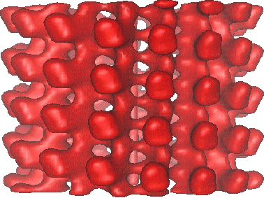
| Title | Nucleotide-induced conformations in the neck region of dimeric kinesin. | |||||||||
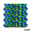
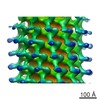
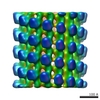
 Map data Map data | rat kinesin Dimer with an SH3 domain cloned within neck region rKS379, complexed to microtubules in the presence of AMP-PNP | |||||||||
 Sample Sample |
| |||||||||
| Biological species |  | |||||||||
| Method | helical reconstruction / cryo EM / negative staining / Resolution: 25.0 Å | |||||||||
 Authors Authors | Skiniotis G | |||||||||
 Citation Citation | Journal: EMBO J / Year: 2003 Title: Nucleotide-induced conformations in the neck region of dimeric kinesin. Authors: Georgios Skiniotis / Thomas Surrey / Stephan Altmann / Heinz Gross / Young-Hwa Song / Eckhard Mandelkow / Andreas Hoenger /  Abstract: The neck region of kinesin constitutes a key component in the enzyme's walking mechanism. Here we applied cryoelectron microscopy and image reconstruction to investigate the location of the kinesin ...The neck region of kinesin constitutes a key component in the enzyme's walking mechanism. Here we applied cryoelectron microscopy and image reconstruction to investigate the location of the kinesin neck in dimeric and monomeric constructs complexed to microtubules. To this end we enhanced the visibility of this region by engineering an SH3 domain into the transition between neck linker and neck coiled coil. The resulting chimeric kinesin constructs remained functional as verified by physiology assays. In the presence of AMP-PNP the SH3 domains allowed us to identify the position of the neck in a well defined conformation and revealed its high flexibility in the absence of nucleotide. We show here the double-headed binding of dimeric kinesin along the same protofilament, which is characterized by the opposite directionality of neck linkers. In this configuration the neck coiled coil appears fully zipped. The position of the neck region in dimeric constructs is not affected by the presence of the tubulin C-termini as confirmed by subtilisin treatment of microtubules prior to motor decoration. | |||||||||
| History |
|
- Structure visualization
| Movie |
 Movie viewer Movie viewer |
|---|---|
| Structure viewer | EM map: SurfViewMolmilJmol/JSmol |
| Supplemental images |
- Downloads & links
-EMDB archive
| Map data | emd_1038.map.gz | 2.9 MB | EMDB map data format | |
|---|---|---|---|---|
| Header (meta data) | emd-1038-v30.xmlemd-1038.xml | 10.5 KB 10.5 KB | Display Display | EMDB header |
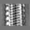
| Images |  1038.gif 1038.gif | 63.5 KB | ||
| Archive directory |  http://ftp.pdbj.org/pub/emdb/structures/EMD-1038ftp://ftp.pdbj.org/pub/emdb/structures/EMD-1038 http://ftp.pdbj.org/pub/emdb/structures/EMD-1038ftp://ftp.pdbj.org/pub/emdb/structures/EMD-1038 | HTTPS FTP |
-Related structure data


-Links
| EMDB pages | EMDB (EBI/PDBe) / EMDataResource |
|---|
-Map
| File | Download / File: emd_1038.map.gz / Format: CCP4 / Size: 3.7 MB / Type: IMAGE STORED AS FLOATING POINT NUMBER (4 BYTES) | ||||||||||||||||||||||||||||||||||||||||||||||||||||||||||||
|---|---|---|---|---|---|---|---|---|---|---|---|---|---|---|---|---|---|---|---|---|---|---|---|---|---|---|---|---|---|---|---|---|---|---|---|---|---|---|---|---|---|---|---|---|---|---|---|---|---|---|---|---|---|---|---|---|---|---|---|---|---|
| Annotation | rat kinesin Dimer with an SH3 domain cloned within neck region rKS379, complexed to microtubules in the presence of AMP-PNP | ||||||||||||||||||||||||||||||||||||||||||||||||||||||||||||
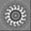
| Projections & slices | Image control
Images are generated by Spider. | ||||||||||||||||||||||||||||||||||||||||||||||||||||||||||||
| Voxel size | X=Y=Z: 5.526 Å | ||||||||||||||||||||||||||||||||||||||||||||||||||||||||||||
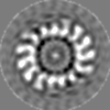
| Density |
| ||||||||||||||||||||||||||||||||||||||||||||||||||||||||||||
| Symmetry | Space group: 1 | ||||||||||||||||||||||||||||||||||||||||||||||||||||||||||||
| Details | EMDB XML:
CCP4 map header:
| ||||||||||||||||||||||||||||||||||||||||||||||||||||||||||||
 Z (Sec.)
 Y (Row.)
 X (Col.)


-Supplemental data
- Sample components
-Entire : rat kinesin construct rK379
| Entire | Name: rat kinesin construct rK379 |
|---|---|
| Components |
|
-Supramolecule #1000: rat kinesin construct rK379
| Supramolecule | Name: rat kinesin construct rK379 / type: sample / ID: 1000 / Oligomeric state: dimer / Number unique components: 2 |
|---|
-Macromolecule #1: rat kinesin
| Macromolecule | Name: rat kinesin / type: protein_or_peptide / ID: 1 / Name.synonym: molecular motor / Number of copies: 1 / Oligomeric state: dimer / Recombinant expression: Yes |
|---|---|
| Source (natural) | Organism: |
| Molecular weight | Experimental: 980 KDa |
| Recombinant expression | Organism:  |
-Macromolecule #2: tubulin
| Macromolecule | Name: tubulin / type: protein_or_peptide / ID: 2 / Name.synonym: microtubule / Number of copies: 1 / Oligomeric state: hetero-dimer / Recombinant expression: No / Database: NCBI |
|---|---|
| Source (natural) | Organism: |
| Molecular weight | Experimental: 110 KDa |
-Experimental details
-Structure determination
| Method | negative staining, cryo EM |
|---|---|
 Processing Processing | helical reconstruction |
| Aggregation state | filament |
-Sample preparation
| Concentration | 0.5 mg/mL |
|---|---|
| Buffer | pH: 6.8 Details: PIPES 80 mM, MgCl2 1 mM, GTP 1 mM, Taxol 20 uM, DMSO 7.5% |
| Staining | Type: NEGATIVE / Details: ice-embeded |
| Grid | Details: holey grids |
| Vitrification | Cryogen name: ETHANE / Chamber temperature: 93 K / Instrument: HOMEMADE PLUNGER / Details: Vitrification instrument: self made |
- Electron microscopy
| Microscope | FEI/PHILIPS CM200FEG/ST |
|---|---|
| Temperature | Average: 95 K |
| Alignment procedure | Legacy - Astigmatism: was corrected at 180,000 times mag. |
| Image recording | Category: FILM / Film or detector model: KODAK SO-163 FILM / Digitization - Scanner: ZEISS SCAI / Digitization - Sampling interval: 21 µm / Number real images: 16 / Average electron dose: 5 e/Å2 / Bits/pixel: 8 |
| Tilt angle min | 0 |
| Tilt angle max | 0 |
| Electron beam | Acceleration voltage: 200 kV / Electron source:  FIELD EMISSION GUN FIELD EMISSION GUN |
| Electron optics | Illumination mode: FLOOD BEAM / Imaging mode: BRIGHT FIELD / Cs: 2 mm / Nominal defocus max: 2.5 µm / Nominal defocus min: 2.0 µm / Nominal magnification: 38000 |
| Sample stage | Specimen holder: side-entry / Specimen holder model: GATAN LIQUID NITROGEN |
-Image processing
| Final reconstruction | Applied symmetry - Helical parameters - Axial symmetry: C1 (asymmetric) Algorithm: OTHER / Resolution.type: BY AUTHOR / Resolution: 25.0 Å / Resolution method: FSC 0.5 CUT-OFF / Software - Name: PHOELIX, SUPREME Details: Final map from 33 averaged datasets = 17 helical tubes |
|---|
