 ムービー
ムービー コントローラー


+ データを開く

- 基本情報
基本情報
| 登録情報 | データベース: EMDB / ID: EMD-1017 | |||||||||
|---|---|---|---|---|---|---|---|---|---|---|
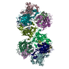
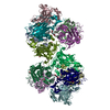
| タイトル | The DnaB.DnaC complex: a structure based on dimers assembled around an occluded channel. | |||||||||
 マップデータ マップデータ | ||||||||||
 試料 試料 |
| |||||||||
| 機能・相同性 | : / DNA helicase activity 機能・相同性情報 機能・相同性情報 | |||||||||
| 生物種 |  | |||||||||
| 手法 | 単粒子再構成法 / クライオ電子顕微鏡法 / 解像度: 26.0 Å | |||||||||
 データ登録者 データ登録者 | Barcena M / Ruiz T / Donate LE / Brown SE / Dixon NE / Radermacher M / Carazo JM | |||||||||
 引用 引用 | ジャーナル: EMBO J / 年: 2001 タイトル: The DnaB.DnaC complex: a structure based on dimers assembled around an occluded channel. 著者: M Bárcena / T Ruiz / L E Donate / S E Brown / N E Dixon / M Radermacher / J M Carazo /  要旨: Replicative helicases are motor proteins that unwind DNA at replication forks. Escherichia coli DnaB is the best characterized member of this family of enzymes. We present the 26 A resolution three- ...Replicative helicases are motor proteins that unwind DNA at replication forks. Escherichia coli DnaB is the best characterized member of this family of enzymes. We present the 26 A resolution three-dimensional structure of the DnaB hexamer in complex with its loading partner, DnaC, obtained from cryo-electron microscopy. Analysis of the volume brings insight into the elaborate way the two proteins interact, and provides a structural basis for control of the symmetry state and inactivation of the helicase by DnaC. The complex is arranged on the basis of interactions among DnaC and DnaB dimers. DnaC monomers are observed for the first time to arrange as three dumb-bell-shaped dimers that interlock into one of the faces of the helicase. This could be responsible for the freezing of DnaB in a C(3) architecture by its loading partner. The central channel of the helicase is almost occluded near the end opposite to DnaC, such that even single-stranded DNA could not pass through. We propose that the DnaB N-terminal domain is located at this face. | |||||||||
| 履歴 |
|
- 構造の表示
構造の表示
| ムービー |
ムービービューア |
|---|---|
| 構造ビューア | EMマップ: SurfViewMolmilJmol/JSmol |
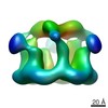
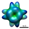
| 添付画像 |
 UCSF Chimera
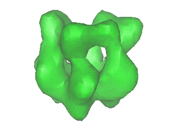
- ダウンロードとリンク
-EMDBアーカイブ
| マップデータ | emd_1017.map.gz | 7.3 MB | EMDBマップデータ形式 | |
|---|---|---|---|---|
| ヘッダ (付随情報) | emd-1017-v30.xmlemd-1017.xml | 9.5 KB 9.5 KB | 表示 表示 | EMDBヘッダ |
| 画像 |  1017.gif 1017.gif | 16.1 KB | ||
| アーカイブディレクトリ |  http://ftp.pdbj.org/pub/emdb/structures/EMD-1017ftp://ftp.pdbj.org/pub/emdb/structures/EMD-1017 http://ftp.pdbj.org/pub/emdb/structures/EMD-1017ftp://ftp.pdbj.org/pub/emdb/structures/EMD-1017 | HTTPS FTP |
-検証レポート
| 文書・要旨 | emd_1017_validation.pdf.gz | 201.6 KB | 表示 | EMDB検証レポート |
|---|---|---|---|---|
| 文書・詳細版 | emd_1017_full_validation.pdf.gz | 200.8 KB | 表示 | |
| XML形式データ | emd_1017_validation.xml.gz | 5.4 KB | 表示 | |
| アーカイブディレクトリ | https://ftp.pdbj.org/pub/emdb/validation_reports/EMD-1017ftp://ftp.pdbj.org/pub/emdb/validation_reports/EMD-1017 | HTTPS FTP |
-関連構造データ

-リンク
| EMDBのページ | EMDB (EBI/PDBe) / EMDataResource |
|---|
-マップ
| ファイル | ダウンロード / ファイル: emd_1017.map.gz / 形式: CCP4 / 大きさ: 7.8 MB / タイプ: IMAGE STORED AS FLOATING POINT NUMBER (4 BYTES) | ||||||||||||||||||||||||||||||||||||||||||||||||||||||||||||
|---|---|---|---|---|---|---|---|---|---|---|---|---|---|---|---|---|---|---|---|---|---|---|---|---|---|---|---|---|---|---|---|---|---|---|---|---|---|---|---|---|---|---|---|---|---|---|---|---|---|---|---|---|---|---|---|---|---|---|---|---|---|
| 投影像・断面図 | 画像のコントロール
画像は Spider により作成 | ||||||||||||||||||||||||||||||||||||||||||||||||||||||||||||
| ボクセルのサイズ | X=Y=Z: 3.6 Å | ||||||||||||||||||||||||||||||||||||||||||||||||||||||||||||
| 密度 |
| ||||||||||||||||||||||||||||||||||||||||||||||||||||||||||||
| 対称性 | 空間群: 1 | ||||||||||||||||||||||||||||||||||||||||||||||||||||||||||||
| 詳細 | EMDB XML:
CCP4マップ ヘッダ情報:
| ||||||||||||||||||||||||||||||||||||||||||||||||||||||||||||
 Z (Sec.)
 Y (Row.)
 X (Col.)


-添付データ
- 試料の構成要素
-全体 : DnaB.DnaC complex from Escherichia coli
| 全体 | 名称: DnaB.DnaC complex from Escherichia coli |
|---|---|
| 要素 |
|
-超分子 #1000: DnaB.DnaC complex from Escherichia coli
| 超分子 | 名称: DnaB.DnaC complex from Escherichia coli / タイプ: sample / ID: 1000 集合状態: one homohexamer of DnaB binds to six monomers of DnaC Number unique components: 2 |
|---|---|
| 分子量 | 理論値: 480 KDa |
-分子 #1: DnaB
| 分子 | 名称: DnaB / タイプ: protein_or_peptide / ID: 1 / コピー数: 6 / 集合状態: hexamer / 組換発現: Yes |
|---|---|
| 由来(天然) | 生物種: |
| 分子量 | 理論値: 310 KDa |
| 組換発現 | 生物種: |
| 配列 | GO: DNA helicase activity / InterPro: INTERPRO: IPR001198 |
-分子 #2: DnaC
| 分子 | 名称: DnaC / タイプ: protein_or_peptide / ID: 2 / コピー数: 6 / 集合状態: three dimers / 組換発現: Yes |
|---|---|
| 由来(天然) | 生物種: |
| 分子量 | 理論値: 170 KDa |
| 組換発現 | 生物種: |
-実験情報
-構造解析
| 手法 | クライオ電子顕微鏡法 |
|---|---|
 解析 解析 | 単粒子再構成法 |
| 試料の集合状態 | particle |
-試料調製
| 濃度 | 0.03 mg/mL |
|---|---|
| 緩衝液 | pH: 7.6 詳細: 50 mM Tris-HCl, 25 mM NaCl 5 mM MgCl2 2 mM DTT 0.1 mM ATP |
| 凍結 | 凍結剤: ETHANE / 装置: HOMEMADE PLUNGER / 詳細: Vitrification instrument: plunger / 手法: double-blotting |
- 電子顕微鏡法
| 顕微鏡 | FEI/PHILIPS CM120T |
|---|---|
| 撮影 | カテゴリ: FILM / フィルム・検出器のモデル: KODAK SO-163 FILM / デジタル化 - スキャナー: ZEISS SCAI / デジタル化 - サンプリング間隔: 7 µm / 実像数: 58 / ビット/ピクセル: 8 |
| Tilt angle min | 0 |
| 電子線 | 加速電圧: 100 kV / 電子線源: LAB6 |
| 電子光学系 | 倍率(補正後): 57874 / 照射モード: FLOOD BEAM / 撮影モード: BRIGHT FIELD / Cs: 2.2 mm / 最大 デフォーカス(公称値): 2.0 µm / 最小 デフォーカス(公称値): 1.0 µm / 倍率(公称値): 60000 |
| 試料ステージ | 試料ホルダー: cryo-holder / 試料ホルダーモデル: GATAN LIQUID NITROGEN / Tilt angle max: 35 |
-画像解析
| 最終 再構成 | 想定した対称性 - 点群: C3 (3回回転対称) / アルゴリズム: OTHER / 解像度のタイプ: BY AUTHOR / 解像度: 26.0 Å / 解像度の算出法: OTHER / ソフトウェア - 名称: SPIDER and Xmipp / 使用した粒子像数: 7888 |
|---|
