 Movie
Movie Controller

[English] 日本語
 Yorodumi
- EMDB-1017: The DnaB.DnaC complex: a structure based on dimers assembled arou... -
+ Open data

- Basic information
| Entry | Database: EMDB / ID: EMD-1017 | |||||||||
|---|---|---|---|---|---|---|---|---|---|---|
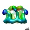
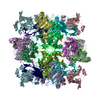
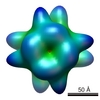
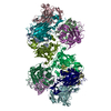
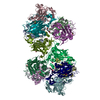
| Title | The DnaB.DnaC complex: a structure based on dimers assembled around an occluded channel. | |||||||||
 Map data Map data | ||||||||||
 Sample Sample |
| |||||||||
| Function / homology | : / DNA helicase activity Function and homology information Function and homology information | |||||||||
| Biological species |  | |||||||||
| Method | single particle reconstruction / cryo EM / Resolution: 26.0 Å | |||||||||
 Authors Authors | Barcena M / Ruiz T / Donate LE / Brown SE / Dixon NE / Radermacher M / Carazo JM | |||||||||
 Citation Citation | Journal: EMBO J / Year: 2001 Title: The DnaB.DnaC complex: a structure based on dimers assembled around an occluded channel. Authors: M Bárcena / T Ruiz / L E Donate / S E Brown / N E Dixon / M Radermacher / J M Carazo /  Abstract: Replicative helicases are motor proteins that unwind DNA at replication forks. Escherichia coli DnaB is the best characterized member of this family of enzymes. We present the 26 A resolution three- ...Replicative helicases are motor proteins that unwind DNA at replication forks. Escherichia coli DnaB is the best characterized member of this family of enzymes. We present the 26 A resolution three-dimensional structure of the DnaB hexamer in complex with its loading partner, DnaC, obtained from cryo-electron microscopy. Analysis of the volume brings insight into the elaborate way the two proteins interact, and provides a structural basis for control of the symmetry state and inactivation of the helicase by DnaC. The complex is arranged on the basis of interactions among DnaC and DnaB dimers. DnaC monomers are observed for the first time to arrange as three dumb-bell-shaped dimers that interlock into one of the faces of the helicase. This could be responsible for the freezing of DnaB in a C(3) architecture by its loading partner. The central channel of the helicase is almost occluded near the end opposite to DnaC, such that even single-stranded DNA could not pass through. We propose that the DnaB N-terminal domain is located at this face. | |||||||||
| History |
|
- Structure visualization
| Movie |
Movie viewer |
|---|---|
| Structure viewer | EM map: SurfViewMolmilJmol/JSmol |
| Supplemental images |
 UCSF Chimera
- Downloads & links
-EMDB archive
| Map data | emd_1017.map.gz | 7.3 MB | EMDB map data format | |
|---|---|---|---|---|
| Header (meta data) | emd-1017-v30.xmlemd-1017.xml | 9.5 KB 9.5 KB | Display Display | EMDB header |
| Images |  1017.gif 1017.gif | 16.1 KB | ||
| Archive directory |  http://ftp.pdbj.org/pub/emdb/structures/EMD-1017ftp://ftp.pdbj.org/pub/emdb/structures/EMD-1017 http://ftp.pdbj.org/pub/emdb/structures/EMD-1017ftp://ftp.pdbj.org/pub/emdb/structures/EMD-1017 | HTTPS FTP |
-Related structure data
| Similar structure data |
|---|

-Links
| EMDB pages | EMDB (EBI/PDBe) / EMDataResource |
|---|
-Map
| File | Download / File: emd_1017.map.gz / Format: CCP4 / Size: 7.8 MB / Type: IMAGE STORED AS FLOATING POINT NUMBER (4 BYTES) | ||||||||||||||||||||||||||||||||||||||||||||||||||||||||||||
|---|---|---|---|---|---|---|---|---|---|---|---|---|---|---|---|---|---|---|---|---|---|---|---|---|---|---|---|---|---|---|---|---|---|---|---|---|---|---|---|---|---|---|---|---|---|---|---|---|---|---|---|---|---|---|---|---|---|---|---|---|---|
| Projections & slices | Image control
Images are generated by Spider. | ||||||||||||||||||||||||||||||||||||||||||||||||||||||||||||
| Voxel size | X=Y=Z: 3.6 Å | ||||||||||||||||||||||||||||||||||||||||||||||||||||||||||||
| Density |
| ||||||||||||||||||||||||||||||||||||||||||||||||||||||||||||
| Symmetry | Space group: 1 | ||||||||||||||||||||||||||||||||||||||||||||||||||||||||||||
| Details | EMDB XML:
CCP4 map header:
| ||||||||||||||||||||||||||||||||||||||||||||||||||||||||||||
 Z (Sec.)
 Y (Row.)
 X (Col.)


-Supplemental data
- Sample components
-Entire : DnaB.DnaC complex from Escherichia coli
| Entire | Name: DnaB.DnaC complex from Escherichia coli |
|---|---|
| Components |
|
-Supramolecule #1000: DnaB.DnaC complex from Escherichia coli
| Supramolecule | Name: DnaB.DnaC complex from Escherichia coli / type: sample / ID: 1000 Oligomeric state: one homohexamer of DnaB binds to six monomers of DnaC Number unique components: 2 |
|---|---|
| Molecular weight | Theoretical: 480 KDa |
-Macromolecule #1: DnaB
| Macromolecule | Name: DnaB / type: protein_or_peptide / ID: 1 / Number of copies: 6 / Oligomeric state: hexamer / Recombinant expression: Yes |
|---|---|
| Source (natural) | Organism: |
| Molecular weight | Theoretical: 310 KDa |
| Recombinant expression | Organism: |
| Sequence | GO: DNA helicase activity / InterPro: INTERPRO: IPR001198 |
-Macromolecule #2: DnaC
| Macromolecule | Name: DnaC / type: protein_or_peptide / ID: 2 / Number of copies: 6 / Oligomeric state: three dimers / Recombinant expression: Yes |
|---|---|
| Source (natural) | Organism: |
| Molecular weight | Theoretical: 170 KDa |
| Recombinant expression | Organism: |
-Experimental details
-Structure determination
| Method | cryo EM |
|---|---|
 Processing Processing | single particle reconstruction |
| Aggregation state | particle |
-Sample preparation
| Concentration | 0.03 mg/mL |
|---|---|
| Buffer | pH: 7.6 Details: 50 mM Tris-HCl, 25 mM NaCl 5 mM MgCl2 2 mM DTT 0.1 mM ATP |
| Vitrification | Cryogen name: ETHANE / Instrument: HOMEMADE PLUNGER / Details: Vitrification instrument: plunger / Method: double-blotting |
- Electron microscopy
| Microscope | FEI/PHILIPS CM120T |
|---|---|
| Image recording | Category: FILM / Film or detector model: KODAK SO-163 FILM / Digitization - Scanner: ZEISS SCAI / Digitization - Sampling interval: 7 µm / Number real images: 58 / Bits/pixel: 8 |
| Tilt angle min | 0 |
| Electron beam | Acceleration voltage: 100 kV / Electron source: LAB6 |
| Electron optics | Calibrated magnification: 57874 / Illumination mode: FLOOD BEAM / Imaging mode: BRIGHT FIELD / Cs: 2.2 mm / Nominal defocus max: 2.0 µm / Nominal defocus min: 1.0 µm / Nominal magnification: 60000 |
| Sample stage | Specimen holder: cryo-holder / Specimen holder model: GATAN LIQUID NITROGEN / Tilt angle max: 35 |
-Image processing
| Final reconstruction | Applied symmetry - Point group: C3 (3 fold cyclic) / Algorithm: OTHER / Resolution.type: BY AUTHOR / Resolution: 26.0 Å / Resolution method: OTHER / Software - Name: SPIDER and Xmipp / Number images used: 7888 |
|---|
