National Health and Medical Research Council (NHMRC, Australia)
1120919
Australia
National Health and Medical Research Council (NHMRC, Australia)
1150083
Australia
National Health and Medical Research Council (NHMRC, Australia)
1159006
Australia
Japan Science and Technology
18069571
Japan
Japan Society for the Promotion of Science (JSPS)
18H06043
Japan
Citation
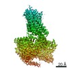
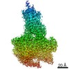
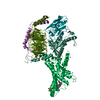
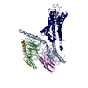
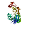
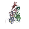
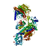
Journal: Mol Cell / Year: 2020 Title: Toward a Structural Understanding of Class B GPCR Peptide Binding and Activation. Authors: Yi-Lynn Liang / Matthew J Belousoff / Peishen Zhao / Cassandra Koole / Madeleine M Fletcher / Tin T Truong / Villy Julita / George Christopoulos / H Eric Xu / Yan Zhang / Maryam Khoshouei / ...Authors: Yi-Lynn Liang / Matthew J Belousoff / Peishen Zhao / Cassandra Koole / Madeleine M Fletcher / Tin T Truong / Villy Julita / George Christopoulos / H Eric Xu / Yan Zhang / Maryam Khoshouei / Arthur Christopoulos / Radostin Danev / Patrick M Sexton / Denise Wootten / Abstract: Class B G protein-coupled receptors (GPCRs) are important therapeutic targets for major diseases. Here, we present structures of peptide and Gs-bound pituitary adenylate cyclase-activating peptide, ...Class B G protein-coupled receptors (GPCRs) are important therapeutic targets for major diseases. Here, we present structures of peptide and Gs-bound pituitary adenylate cyclase-activating peptide, PAC1 receptor, and corticotropin-releasing factor (CRF), (CRF1) receptor. Together with recently solved structures, these provide coverage of the major class B GPCR subfamilies. Diverse orientations of the extracellular domain to the receptor core in different receptors are at least partially dependent on evolutionary conservation in the structure and nature of peptide interactions. Differences in peptide interactions to the receptor core also influence the interlinked TM2-TM1-TM6/ECL3/TM7 domain, and this is likely important in their diverse signaling. However, common conformational reorganization of ECL2, linked to reorganization of ICL2, modulates G protein contacts. Comparison between receptors reveals ICL2 as a key domain forming dynamic G protein interactions in a receptor- and ligand-specific manner. This work advances our understanding of class B GPCR activation and Gs coupling.
History
Deposition
Feb 3, 2020
-
Header (metadata) release
Feb 19, 2020
-
Map release
Feb 19, 2020
-
Update
Dec 9, 2020
-
Current status
Dec 9, 2020
Processing site: PDBj / Status: Released
-
Structure visualization
Movie
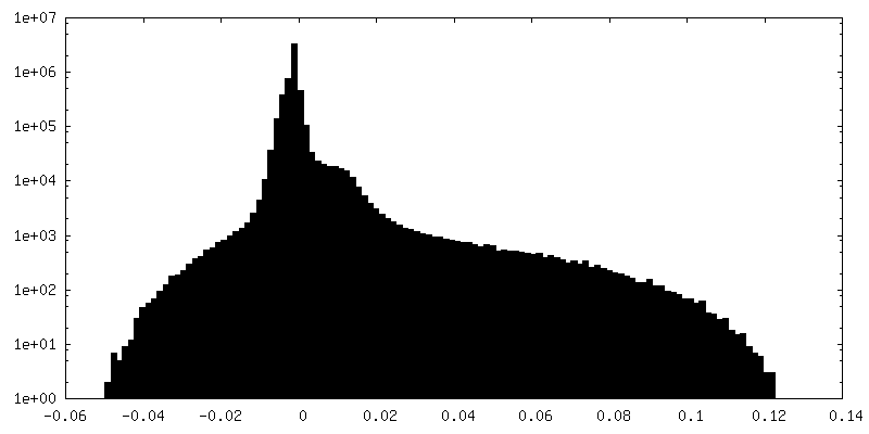
Surface view with section colored by density value
EMPIAR-10359 (Title: Cryo-EM of PAC1 receptor bound to PACAP38 and Gs protein Data size: 4.8 TB Data #1: Unaligned multi-frame gain-normalized movies in LZW compressed TIFF format [micrographs - multiframe])
Cryogen name: ETHANE / Chamber humidity: 100 % / Chamber temperature: 277 K / Instrument: FEI VITROBOT MARK IV / Details: blot time 10 s.
-
Electron microscopy
Microscope
FEI TITAN KRIOS
Specialist optics
Phase plate: VOLTA PHASE PLATE / Energy filter - Name: GIF Quantum LS / Energy filter - Slit width: 25 eV
Image recording
Film or detector model: GATAN K3 BIOQUANTUM (6k x 4k) / Number grids imaged: 1 / Number real images: 7650 / Average exposure time: 3.715 sec. / Average electron dose: 64.0 e/Å2 Details: Volta phase plate images: 4032; Conventional defocus images: 3618
Electron beam
Acceleration voltage: 300 kV / Electron source: FIELD EMISSION GUN
In the structure databanks used in Yorodumi, some data are registered as the other names, "COVID-19 virus" and "2019-nCoV". Here are the details of the virus and the list of structure data.
Jan 31, 2019. EMDB accession codes are about to change! (news from PDBe EMDB page)
EMDB accession codes are about to change! (news from PDBe EMDB page)
The allocation of 4 digits for EMDB accession codes will soon come to an end. Whilst these codes will remain in use, new EMDB accession codes will include an additional digit and will expand incrementally as the available range of codes is exhausted. The current 4-digit format prefixed with “EMD-” (i.e. EMD-XXXX) will advance to a 5-digit format (i.e. EMD-XXXXX), and so on. It is currently estimated that the 4-digit codes will be depleted around Spring 2019, at which point the 5-digit format will come into force.
The EM Navigator/Yorodumi systems omit the EMD- prefix.
Related info.:Q: What is EMD? / ID/Accession-code notation in Yorodumi/EM Navigator
Yorodumi is a browser for structure data from EMDB, PDB, SASBDB, etc.
This page is also the successor to EM Navigator detail page, and also detail information page/front-end page for Omokage search.
The word "yorodu" (or yorozu) is an old Japanese word meaning "ten thousand". "mi" (miru) is to see.
Related info.:EMDB / PDB / SASBDB / Comparison of 3 databanks / Yorodumi Search / Aug 31, 2016. New EM Navigator & Yorodumi / Yorodumi Papers / Jmol/JSmol / Function and homology information / Changes in new EM Navigator and Yorodumi
 Movie
Movie Controller


 Open data
 Basic information
 Map data
 Sample
 Function and homology information
 Homo sapiens (human)
 Authors
 Australia,
  Japan, 5 items
  Citation


 Structure visualization
 Downloads & links
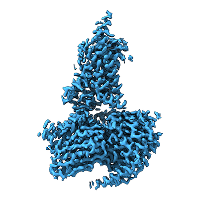 emd_0993.png
 http://ftp.pdbj.org/pub/emdb/structures/EMD-0993


 Z (Sec.)
 Y (Row.)
 X (Col.)


 Sample components
 Trichoplusia ni (cabbage looper)
 Processing
 Electron microscopy
 FIELD EMISSION GUN
