 Movie
Movie Controller


+ Open data

- Basic information
| Entry | Database: EMDB / ID: EMD-0938 | |||||||||
|---|---|---|---|---|---|---|---|---|---|---|
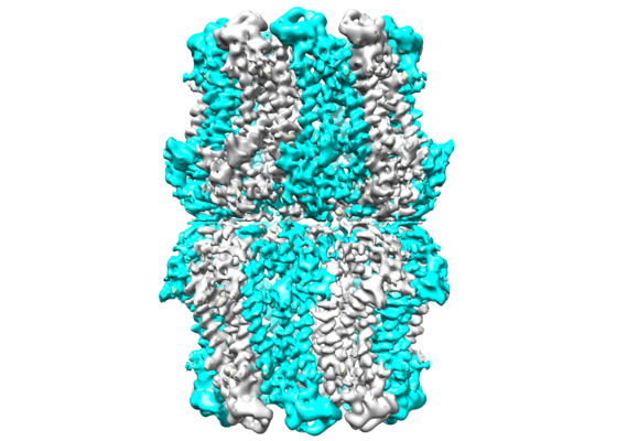
| Title | Structure of CLHM1 from Caenorhabditis Elegans | |||||||||
 Map data Map data | ||||||||||
 Sample Sample |
| |||||||||
 Keywords Keywords | complex / MEMBRANE PROTEIN | |||||||||
| Function / homology |  Function and homology information Function and homology informationATP export / regulation of locomotion / non-motile cilium / monoatomic ion channel complex / voltage-gated calcium channel activity / monoatomic cation channel activity / calcium ion transmembrane transport / plasma membrane Similarity search - Function | |||||||||
| Biological species |  | |||||||||
| Method | single particle reconstruction / cryo EM / Resolution: 3.73 Å | |||||||||
 Authors Authors | Yang WX / Wang YW | |||||||||
| Funding support |  China, 1 items China, 1 items
| |||||||||
 Citation Citation | Journal: Protein Sci / Year: 2020 Title: Cryo-electron microscopy structure of CLHM1 ion channel from Caenorhabditis elegans. Authors: Weixin Yang / Youwang Wang / Jianli Guo / Lingli He / Ye Zhou / Hui Zheng / Zhenfeng Liu / Ping Zhu / Xuejun C Zhang / Abstract: Calcium homeostasis modulators (CALHMs/CLHMs) comprise a family of pore-forming protein complexes assembling into voltage-gated, Ca -sensitive, nonselective channels. These complexes contain an ion- ...Calcium homeostasis modulators (CALHMs/CLHMs) comprise a family of pore-forming protein complexes assembling into voltage-gated, Ca -sensitive, nonselective channels. These complexes contain an ion-conduction pore sufficiently wide to permit the passing of ATP molecules serving as neurotransmitters. While their function and structure information is accumulating, the precise mechanisms of these channel complexes remain to be full understood. Here, we present the structure of the Caenorhabditis elegans CLHM1 channel in its open state solved through single-particle cryo-electron microscopy at 3.7-Å resolution. The transmembrane region of the channel structure of the dominant class shows an assembly of 10-fold rotational symmetry in one layer, and its cytoplasmic region is involved in additional twofold symmetrical packing in a tail-to-tail manner. Furthermore, we identified a series of amino acid residues critical for the regulation of CeCLHM1 channel using functional assays, electrophysiological analyses as well as structural-based analysis. Our structure and function analyses provide new insights into the mechanisms of CALHM channels. | |||||||||
| History |
|
- Structure visualization
| Movie |
Movie viewer |
|---|---|
| Structure viewer | EM map: SurfViewMolmilJmol/JSmol |
| Supplemental images |
- Downloads & links
-EMDB archive
| Map data | emd_0938.map.gz | 12 MB | EMDB map data format | |
|---|---|---|---|---|
| Header (meta data) | emd-0938-v30.xmlemd-0938.xml | 9 KB 9 KB | Display Display | EMDB header |
| Images |  emd_0938.png emd_0938.png | 217.8 KB | ||
| Filedesc metadata | emd-0938.cif.gz | 4.9 KB | ||
| Archive directory |  http://ftp.pdbj.org/pub/emdb/structures/EMD-0938ftp://ftp.pdbj.org/pub/emdb/structures/EMD-0938 http://ftp.pdbj.org/pub/emdb/structures/EMD-0938ftp://ftp.pdbj.org/pub/emdb/structures/EMD-0938 | HTTPS FTP |
-Related structure data
| Related structure data |  6lomMC M: atomic model generated by this map C: citing same article ( |
|---|---|
| Similar structure data |

-Links
| EMDB pages | EMDB (EBI/PDBe) / EMDataResource |
|---|
-Map
| File | Download / File: emd_0938.map.gz / Format: CCP4 / Size: 103 MB / Type: IMAGE STORED AS FLOATING POINT NUMBER (4 BYTES) | ||||||||||||||||||||||||||||||||||||||||||||||||||||||||||||
|---|---|---|---|---|---|---|---|---|---|---|---|---|---|---|---|---|---|---|---|---|---|---|---|---|---|---|---|---|---|---|---|---|---|---|---|---|---|---|---|---|---|---|---|---|---|---|---|---|---|---|---|---|---|---|---|---|---|---|---|---|---|
| Projections & slices | Image control
Images are generated by Spider. | ||||||||||||||||||||||||||||||||||||||||||||||||||||||||||||
| Voxel size | X=Y=Z: 1 Å | ||||||||||||||||||||||||||||||||||||||||||||||||||||||||||||
| Density |
| ||||||||||||||||||||||||||||||||||||||||||||||||||||||||||||
| Symmetry | Space group: 1 | ||||||||||||||||||||||||||||||||||||||||||||||||||||||||||||
| Details | EMDB XML:
CCP4 map header:
| ||||||||||||||||||||||||||||||||||||||||||||||||||||||||||||
 Z (Sec.)
 Y (Row.)
 X (Col.)


-Supplemental data
- Sample components
-Entire : predicted: CALHM1-like protein
| Entire | Name: predicted: CALHM1-like protein |
|---|---|
| Components |
|
-Supramolecule #1: predicted: CALHM1-like protein
| Supramolecule | Name: predicted: CALHM1-like protein / type: complex / ID: 1 / Parent: 0 / Macromolecule list: all |
|---|---|
| Source (natural) | Organism: |
-Macromolecule #1: Calcium homeostasis modulator protein
| Macromolecule | Name: Calcium homeostasis modulator protein / type: protein_or_peptide / ID: 1 / Number of copies: 20 / Enantiomer: LEVO |
|---|---|
| Source (natural) | Organism: |
| Molecular weight | Theoretical: 37.308633 KDa |
| Recombinant expression | Organism:  Homo sapiens (human) Homo sapiens (human) |
| Sequence | String: MTTSINSVVT VFQNVFTNHG STLLNGILIA TTVGGQSLVR KLTFSCPCAY PLNIYHSLVF MFGPTAALLL IGITVNSTTW KLAHGFFFR VRDTRHSWKT TCVSWIEVLI QSSVAPIAWL FVVFLDGGYY RCYRSHEFCL ISDAILCKNS TILNSYASTS S FNKISDNG ...String: MTTSINSVVT VFQNVFTNHG STLLNGILIA TTVGGQSLVR KLTFSCPCAY PLNIYHSLVF MFGPTAALLL IGITVNSTTW KLAHGFFFR VRDTRHSWKT TCVSWIEVLI QSSVAPIAWL FVVFLDGGYY RCYRSHEFCL ISDAILCKNS TILNSYASTS S FNKISDNG KYCPPCICVP NPTDASYLEA ESQIYAWGLL LFSGVAAFLV ITCNRMCDKY TLVQRQYVET YKNVETQKFD AV AKEHASQ LAEHNARAFF GQKDWTKRDW DWVSGIPEVN NPLFARLRLI AAEKTQQTMY TPLQLWNDNK GYRIPQPDLQ LTQ IIVDET KED UniProtKB: Calcium homeostasis modulator protein |
-Experimental details
-Structure determination
| Method | cryo EM |
|---|---|
 Processing Processing | single particle reconstruction |
| Aggregation state | particle |
-Sample preparation
| Buffer | pH: 7.4 |
|---|---|
| Vitrification | Cryogen name: ETHANE |
- Electron microscopy
| Microscope | FEI TALOS ARCTICA |
|---|---|
| Image recording | Film or detector model: GATAN K2 SUMMIT (4k x 4k) / Average electron dose: 60.0 e/Å2 |
| Electron beam | Acceleration voltage: 200 kV / Electron source:  FIELD EMISSION GUN FIELD EMISSION GUN |
| Electron optics | Illumination mode: FLOOD BEAM / Imaging mode: BRIGHT FIELD |
| Experimental equipment |  Model: Talos Arctica / Image courtesy: FEI Company |
-Image processing
| Startup model | Type of model: OTHER |
|---|---|
| Final reconstruction | Resolution.type: BY AUTHOR / Resolution: 3.73 Å / Resolution method: FSC 0.143 CUT-OFF / Number images used: 49000 |
| Initial angle assignment | Type: MAXIMUM LIKELIHOOD |
| Final angle assignment | Type: MAXIMUM LIKELIHOOD |
