 Movie
Movie Controller

[English] 日本語
 Yorodumi
- EMDB-0934: Sub-tomogram averaging of immature Zika virus in complex with Fab... -
+ Open data

- Basic information
| Entry | Database: EMDB / ID: EMD-0934 | ||||||||||||
|---|---|---|---|---|---|---|---|---|---|---|---|---|---|
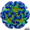
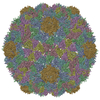
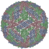
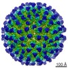
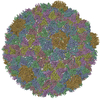
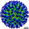
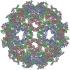
| Title | Sub-tomogram averaging of immature Zika virus in complex with Fab DV62.5 | ||||||||||||
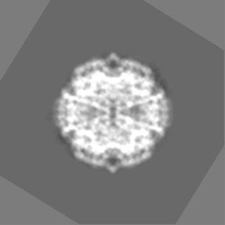
 Map data Map data | |||||||||||||
 Sample Sample |
| ||||||||||||
| Biological species |  Zika virus ZIKV/H. sapiens/FrenchPolynesia/10087PF/2013 / Zika virus ZIKV/H. sapiens/FrenchPolynesia/10087PF/2013 /  Homo sapiens (human) Homo sapiens (human) | ||||||||||||
| Method | subtomogram averaging / cryo EM / Resolution: 13.0 Å | ||||||||||||
 Authors Authors | Tan TY / Fibriansah G / Kostyuchenko VA / Ng TS / Lim XX / Lim XN / Shi J / Morais MC / Corti D / Lok SM | ||||||||||||
| Funding support |  Singapore, 3 items Singapore, 3 items
| ||||||||||||
 Citation Citation | Journal: Nat Commun / Year: 2020 Title: Capsid protein structure in Zika virus reveals the flavivirus assembly process. Authors: Ter Yong Tan / Guntur Fibriansah / Victor A Kostyuchenko / Thiam-Seng Ng / Xin-Xiang Lim / Shuijun Zhang / Xin-Ni Lim / Jiaqi Wang / Jian Shi / Marc C Morais / Davide Corti / Shee-Mei Lok /   Abstract: Structures of flavivirus (dengue virus and Zika virus) particles are known to near-atomic resolution and show detailed structure and arrangement of their surface proteins (E and prM in immature virus ...Structures of flavivirus (dengue virus and Zika virus) particles are known to near-atomic resolution and show detailed structure and arrangement of their surface proteins (E and prM in immature virus or M in mature virus). By contrast, the arrangement of the capsid proteins:RNA complex, which forms the core of the particle, is poorly understood, likely due to inherent dynamics. Here, we stabilize immature Zika virus via an antibody that binds across the E and prM proteins, resulting in a subnanometer resolution structure of capsid proteins within the virus particle. Fitting of the capsid protein into densities shows the presence of a helix previously thought to be removed via proteolysis. This structure illuminates capsid protein quaternary organization, including its orientation relative to the lipid membrane and the genomic RNA, and its interactions with the transmembrane regions of the surface proteins. Results show the capsid protein plays a central role in the flavivirus assembly process. | ||||||||||||
| History |
|
- Structure visualization
| Movie |
 Movie viewer Movie viewer |
|---|---|
| Structure viewer | EM map: SurfViewMolmilJmol/JSmol |
| Supplemental images |

- Downloads & links
-EMDB archive
| Map data | emd_0934.map.gz | 9.8 MB | EMDB map data format | |
|---|---|---|---|---|
| Header (meta data) | emd-0934-v30.xmlemd-0934.xml | 13.2 KB 13.2 KB | Display Display | EMDB header |
| Images |  emd_0934.png emd_0934.png | 194.8 KB | ||
| Archive directory |  http://ftp.pdbj.org/pub/emdb/structures/EMD-0934ftp://ftp.pdbj.org/pub/emdb/structures/EMD-0934 http://ftp.pdbj.org/pub/emdb/structures/EMD-0934ftp://ftp.pdbj.org/pub/emdb/structures/EMD-0934 | HTTPS FTP |
-Related structure data


-Links
| EMDB pages | EMDB (EBI/PDBe) / EMDataResource |
|---|---|
| Related items in Molecule of the Month |

-Map
| File | Download / File: emd_0934.map.gz / Format: CCP4 / Size: 125 MB / Type: IMAGE STORED AS FLOATING POINT NUMBER (4 BYTES) | ||||||||||||||||||||||||||||||||||||||||||||||||||||||||||||
|---|---|---|---|---|---|---|---|---|---|---|---|---|---|---|---|---|---|---|---|---|---|---|---|---|---|---|---|---|---|---|---|---|---|---|---|---|---|---|---|---|---|---|---|---|---|---|---|---|---|---|---|---|---|---|---|---|---|---|---|---|---|
| Projections & slices | Image control
Images are generated by Spider. | ||||||||||||||||||||||||||||||||||||||||||||||||||||||||||||
| Voxel size | X=Y=Z: 3.42 Å | ||||||||||||||||||||||||||||||||||||||||||||||||||||||||||||
| Density |
| ||||||||||||||||||||||||||||||||||||||||||||||||||||||||||||
| Symmetry | Space group: 1 | ||||||||||||||||||||||||||||||||||||||||||||||||||||||||||||
| Details | EMDB XML:
CCP4 map header:
| ||||||||||||||||||||||||||||||||||||||||||||||||||||||||||||
 Z (Sec.)
 Y (Row.)
 X (Col.)


-Supplemental data
- Sample components
-Entire : Zika virus ZIKV/H. sapiens/FrenchPolynesia/10087PF/2013
| Entire | Name: Zika virus ZIKV/H. sapiens/FrenchPolynesia/10087PF/2013 |
|---|---|
| Components |
|
-Supramolecule #1: Zika virus ZIKV/H. sapiens/FrenchPolynesia/10087PF/2013
| Supramolecule | Name: Zika virus ZIKV/H. sapiens/FrenchPolynesia/10087PF/2013 type: virus / ID: 1 / Parent: 0 / Macromolecule list: #1-#5 Details: The virus was isolated from Zika patient. The immature Zika virus was grown in Aedes Albopictus clone C6/36 cell. The anti-prM antibody DV62.5 was generated from EBV-immortalized PBMC that ...Details: The virus was isolated from Zika patient. The immature Zika virus was grown in Aedes Albopictus clone C6/36 cell. The anti-prM antibody DV62.5 was generated from EBV-immortalized PBMC that was obtained from dengue patient. Virus type: VIRION / Virus isolate: STRAIN / Virus enveloped: Yes / Virus empty: No |
|---|
-Supramolecule #2: Zika virus
| Supramolecule | Name: Zika virus / type: complex / ID: 2 / Parent: 1 / Macromolecule list: #1-#2, #5 |
|---|---|
| Source (natural) | Organism: Zika virus ZIKV/H. sapiens/FrenchPolynesia/10087PF/2013 Strain: H/PF/2013 |
-Supramolecule #3: anti-prM antibody DV62.5
| Supramolecule | Name: anti-prM antibody DV62.5 / type: complex / ID: 3 / Parent: 1 / Macromolecule list: #3-#4 |
|---|---|
| Source (natural) | Organism: Homo sapiens (human) |
| Recombinant expression | Organism: Human herpesvirus 4 (strain B95-8) (Epstein-Barr virus (strain B95-8)) |
-Experimental details
-Structure determination
| Method | cryo EM |
|---|---|
 Processing Processing | subtomogram averaging |
| Aggregation state | particle |
-Sample preparation
| Buffer | pH: 8 / Details: 10 mM Tris-HCl, 120 mM NaCl, 1 mM EDTA, pH 8 |
|---|---|
| Vitrification | Cryogen name: ETHANE |
| Details | Immature Zika virus-Fab DV62.5 complex sample was prepared by mixing purified immature Zika virus with Fab DV62.5 at an E protein-Fab DV62.5 ratio of 1:1.1 and then the mixture was incubated at 37deg C for 30 min. The sample then was mixed with solution of 10 nm gold particles. |
- Electron microscopy
| Microscope | FEI TITAN KRIOS |
|---|---|
| Image recording | Film or detector model: FEI FALCON II (4k x 4k) / Average electron dose: 3.0 e/Å2 |
| Electron beam | Acceleration voltage: 300 kV / Electron source:  FIELD EMISSION GUN FIELD EMISSION GUN |
| Electron optics | Illumination mode: SPOT SCAN / Imaging mode: BRIGHT FIELD / Cs: 2.7 mm / Nominal defocus max: 4.0 µm / Nominal defocus min: 3.0 µm / Nominal magnification: 47000 |
| Experimental equipment |  Model: Titan Krios / Image courtesy: FEI Company |
-Image processing
| Final reconstruction | Applied symmetry - Point group: C1 (asymmetric) / Resolution.type: BY AUTHOR / Resolution: 13.0 Å / Resolution method: FSC 0.143 CUT-OFF / Software - Name: emClarity / Number subtomograms used: 1152 |
|---|---|
| Extraction | Number tomograms: 63 / Number images used: 1934 / Software - Name: emClarity |
| CTF correction | Software - Name: emClarity |
| Final angle assignment | Type: OTHER / Software - Name: emClarity (ver. 2.1) Details: a missing-wedge constrained cross correlation grid search |
-Atomic model buiding 1
| Refinement | Protocol: FLEXIBLE FIT |
|---|
