 Movie
Movie Controller

[English] 日本語
 Yorodumi
- EMDB-0803: Cryo-EM structure of phosphorylated Tyr39 alpha-synuclein amyloid... -
+ Open data

- Basic information
| Entry | Database: EMDB / ID: EMD-0803 | |||||||||
|---|---|---|---|---|---|---|---|---|---|---|
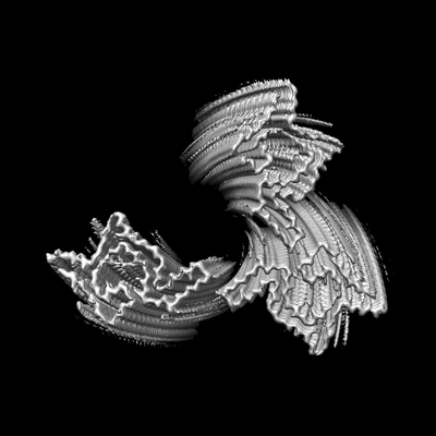
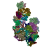
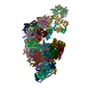
| Title | Cryo-EM structure of phosphorylated Tyr39 alpha-synuclein amyloid fibril | |||||||||
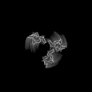
 Map data Map data | ||||||||||
 Sample Sample |
| |||||||||
 Keywords Keywords | Amyloid fibril / PROTEIN FIBRIL | |||||||||
| Function / homology |  Function and homology information Function and homology informationnegative regulation of mitochondrial electron transport, NADH to ubiquinone / neutral lipid metabolic process / regulation of acyl-CoA biosynthetic process / negative regulation of dopamine uptake involved in synaptic transmission / negative regulation of norepinephrine uptake / response to desipramine / positive regulation of SNARE complex assembly / positive regulation of hydrogen peroxide catabolic process / supramolecular fiber / regulation of synaptic vesicle recycling ...negative regulation of mitochondrial electron transport, NADH to ubiquinone / neutral lipid metabolic process / regulation of acyl-CoA biosynthetic process / negative regulation of dopamine uptake involved in synaptic transmission / negative regulation of norepinephrine uptake / response to desipramine / positive regulation of SNARE complex assembly / positive regulation of hydrogen peroxide catabolic process / supramolecular fiber / regulation of synaptic vesicle recycling / negative regulation of chaperone-mediated autophagy / regulation of reactive oxygen species biosynthetic process / positive regulation of protein localization to cell periphery / mitochondrial membrane organization / negative regulation of exocytosis / regulation of glutamate secretion / dopamine biosynthetic process / regulation of macrophage activation / positive regulation of neurotransmitter secretion / response to iron(II) ion / negative regulation of dopamine metabolic process / negative regulation of platelet-derived growth factor receptor signaling pathway / SNARE complex assembly / negative regulation of thrombin-activated receptor signaling pathway / Lewy body / regulation of locomotion / negative regulation of microtubule polymerization / synaptic vesicle priming / regulation of norepinephrine uptake / transporter regulator activity / protein kinase inhibitor activity / positive regulation of inositol phosphate biosynthetic process / synaptic vesicle transport / dopamine uptake involved in synaptic transmission / regulation of dopamine secretion / positive regulation of receptor recycling / cuprous ion binding / positive regulation of exocytosis / nuclear outer membrane / mitochondrial ATP synthesis coupled electron transport / dynein complex binding / synaptic vesicle exocytosis / response to magnesium ion / positive regulation of endocytosis / negative regulation of serotonin uptake / response to type II interferon / cysteine-type endopeptidase inhibitor activity / kinesin binding / regulation of presynapse assembly / synaptic vesicle endocytosis / alpha-tubulin binding / beta-tubulin binding / phospholipase binding / phospholipid metabolic process / supramolecular fiber organization / behavioral response to cocaine / cellular response to fibroblast growth factor stimulus / cellular response to epinephrine stimulus / inclusion body / Hsp70 protein binding / enzyme inhibitor activity / response to interleukin-1 / axon terminus / cellular response to copper ion / regulation of microtubule cytoskeleton organization / positive regulation of release of sequestered calcium ion into cytosol / SNARE binding / adult locomotory behavior / glutathione metabolic process / protein tetramerization / protein sequestering activity / phosphoprotein binding / excitatory postsynaptic potential / tubulin binding / microglial cell activation / ferrous iron binding / fatty acid metabolic process / phospholipid binding / PKR-mediated signaling / synapse organization / receptor internalization / regulation of long-term neuronal synaptic plasticity / protein destabilization / tau protein binding / enzyme activator activity / positive regulation of inflammatory response / terminal bouton / long-term synaptic potentiation / actin cytoskeleton / synaptic vesicle membrane / growth cone / actin binding / cellular response to oxidative stress / neuron apoptotic process / cell cortex / histone binding / response to lipopolysaccharide / microtubule binding / amyloid fibril formation / chemical synaptic transmission Similarity search - Function | |||||||||
| Biological species |  Homo sapiens (human) Homo sapiens (human) | |||||||||
| Method | helical reconstruction / cryo EM / Resolution: 3.37 Å | |||||||||
 Authors Authors | Liu C / Li YM | |||||||||
 Citation Citation | Journal: Proc Natl Acad Sci U S A / Year: 2020 Title: Parkinson's disease-related phosphorylation at Tyr39 rearranges α-synuclein amyloid fibril structure revealed by cryo-EM. Authors: Kun Zhao / Yeh-Jun Lim / Zhenying Liu / Houfang Long / Yunpeng Sun / Jin-Jian Hu / Chunyu Zhao / Youqi Tao / Xing Zhang / Dan Li / Yan-Mei Li / Cong Liu /  Abstract: Posttranslational modifications (PTMs) of α-synuclein (α-syn), e.g., phosphorylation, play an important role in modulating α-syn pathology in Parkinson's disease (PD) and α-synucleinopathies. ...Posttranslational modifications (PTMs) of α-synuclein (α-syn), e.g., phosphorylation, play an important role in modulating α-syn pathology in Parkinson's disease (PD) and α-synucleinopathies. Accumulation of phosphorylated α-syn fibrils in Lewy bodies and Lewy neurites is the histological hallmark of these diseases. However, it is unclear how phosphorylation relates to α-syn pathology. Here, by combining chemical synthesis and bacterial expression, we obtained homogeneous α-syn fibrils with site-specific phosphorylation at Y39, which exhibits enhanced neuronal pathology in rat primary cortical neurons. We determined the cryo-electron microscopy (cryo-EM) structure of the pY39 α-syn fibril, which reveals a fold of α-syn with pY39 in the center of the fibril core forming an electrostatic interaction network with eight charged residues in the N-terminal region of α-syn. This structure composed of residues 1 to 100 represents the largest α-syn fibril core determined so far. This work provides structural understanding on the pathology of the pY39 α-syn fibril and highlights the importance of PTMs in defining the polymorphism and pathology of amyloid fibrils in neurodegenerative diseases. | |||||||||
| History |
|
- Structure visualization
| Movie |
Movie viewer |
|---|---|
| Structure viewer | EM map: SurfViewMolmilJmol/JSmol |
| Supplemental images |
- Downloads & links
-EMDB archive
| Map data | emd_0803.map.gz | 16.7 MB | EMDB map data format | |
|---|---|---|---|---|
| Header (meta data) | emd-0803-v30.xmlemd-0803.xml | 9.1 KB 9.1 KB | Display Display | EMDB header |
| FSC (resolution estimation) | emd_0803_fsc.xml | 11.3 KB | Display | FSC data file |
| Images |  emd_0803.png emd_0803.png | 42 KB | ||
| Filedesc metadata | emd-0803.cif.gz | 4.4 KB | ||
| Archive directory |  http://ftp.pdbj.org/pub/emdb/structures/EMD-0803ftp://ftp.pdbj.org/pub/emdb/structures/EMD-0803 http://ftp.pdbj.org/pub/emdb/structures/EMD-0803ftp://ftp.pdbj.org/pub/emdb/structures/EMD-0803 | HTTPS FTP |
-Related structure data
| Related structure data |  6l1uMC  0801C  6l1tC C: citing same article ( M: atomic model generated by this map |
|---|---|
| Similar structure data |


-Links
| EMDB pages | EMDB (EBI/PDBe) / EMDataResource |
|---|---|
| Related items in Molecule of the Month |

-Map
| File | Download / File: emd_0803.map.gz / Format: CCP4 / Size: 125 MB / Type: IMAGE STORED AS FLOATING POINT NUMBER (4 BYTES) | ||||||||||||||||||||||||||||||||||||||||||||||||||||||||||||
|---|---|---|---|---|---|---|---|---|---|---|---|---|---|---|---|---|---|---|---|---|---|---|---|---|---|---|---|---|---|---|---|---|---|---|---|---|---|---|---|---|---|---|---|---|---|---|---|---|---|---|---|---|---|---|---|---|---|---|---|---|---|
| Projections & slices | Image control
Images are generated by Spider. | ||||||||||||||||||||||||||||||||||||||||||||||||||||||||||||
| Voxel size | X=Y=Z: 1.307 Å | ||||||||||||||||||||||||||||||||||||||||||||||||||||||||||||
| Density |
| ||||||||||||||||||||||||||||||||||||||||||||||||||||||||||||
| Symmetry | Space group: 1 | ||||||||||||||||||||||||||||||||||||||||||||||||||||||||||||
| Details | EMDB XML:
CCP4 map header:
| ||||||||||||||||||||||||||||||||||||||||||||||||||||||||||||
 Z (Sec.)
 Y (Row.)
 X (Col.)


-Supplemental data
- Sample components
-Entire : phosphorylated Tyr39 a-synuclein amyloid fibril
| Entire | Name: phosphorylated Tyr39 a-synuclein amyloid fibril |
|---|---|
| Components |
|
-Supramolecule #1: phosphorylated Tyr39 a-synuclein amyloid fibril
| Supramolecule | Name: phosphorylated Tyr39 a-synuclein amyloid fibril / type: organelle_or_cellular_component / ID: 1 / Parent: 0 / Macromolecule list: all |
|---|---|
| Source (natural) | Organism: Homo sapiens (human) |
-Macromolecule #1: Alpha-synuclein
| Macromolecule | Name: Alpha-synuclein / type: protein_or_peptide / ID: 1 / Number of copies: 15 / Enantiomer: LEVO |
|---|---|
| Source (natural) | Organism: Homo sapiens (human) |
| Molecular weight | Theoretical: 14.556087 KDa |
| Sequence | String: MDVFMKGLSK AKEGVVAAAE KTKQGVAEAA GKTKEGVL(PTR)V GSKTKEGVVH GVATVAEKTK EQVTNVGGAV VTGVTA VAQ KTVEGAGSIA AATGFVKKDQ LGKNEEGAPQ EGILEDMPVD PDNEAYEMPS EEGYQDYEPE A UniProtKB: Alpha-synuclein |
-Experimental details
-Structure determination
| Method | cryo EM |
|---|---|
 Processing Processing | helical reconstruction |
| Aggregation state | filament |
-Sample preparation
| Buffer | pH: 7.5 |
|---|---|
| Vitrification | Cryogen name: ETHANE |
- Electron microscopy
| Microscope | FEI TITAN KRIOS |
|---|---|
| Image recording | Film or detector model: GATAN K2 SUMMIT (4k x 4k) / Average electron dose: 49.0 e/Å2 |
| Electron beam | Acceleration voltage: 300 kV / Electron source:  FIELD EMISSION GUN FIELD EMISSION GUN |
| Electron optics | Illumination mode: FLOOD BEAM / Imaging mode: BRIGHT FIELD |
| Experimental equipment |  Model: Titan Krios / Image courtesy: FEI Company |
-Image processing
| Final reconstruction | Applied symmetry - Helical parameters - Δz: 4.8035 Å Applied symmetry - Helical parameters - Δ&Phi: -0.690334 ° Applied symmetry - Helical parameters - Axial symmetry: C1 (asymmetric) Resolution.type: BY AUTHOR / Resolution: 3.37 Å / Resolution method: FSC 0.143 CUT-OFF / Number images used: 14178 |
|---|---|
| Startup model | Type of model: PDB ENTRY |
| Final angle assignment | Type: NOT APPLICABLE |
| FSC plot (resolution estimation) |  |
